

Translate this page into:
A young lady with abdominal pain and facial lesions
This is an open-access article distributed under the terms of the Creative Commons Attribution License, which permits unrestricted use, distribution, and reproduction in any medium, provided the original work is properly cited.
This article was originally published by Medknow Publications and was migrated to Scientific Scholar after the change of Publisher.
A 28-year-old female presented with abdominal pain for three months, which was constant with no specific character or aggravating factors. Her past was unremarkable except for multiple facial cherry red spots which were asymptomatic. She had completed high school and never had seizures. None of her family members had similar facial lesions. Physical examination revealed red colored papules over the face [Figure 1]. Rest of the examination was normal. Her baseline labs including renal function tests were normal. Computerized tomography (CT) of the abdomen showed a mass lesion in the upper pole of the left kidney with fat attenuation which was suggestive of angiomyolipoma [Figure 2]. CT-brain and echocardiography was normal. A diagnosis of tuberous sclerosis without neurological involvement complicated by symptomatic renal angiomyolipoma was made. In view of persistent abdominal pain the angiomyolipoma was treated with selective renal artery embolization. Her abdominal pain resolved in a week’s time. Follow-up screening, CT after eight weeks showed complete resolution of the angiomyolipoma.
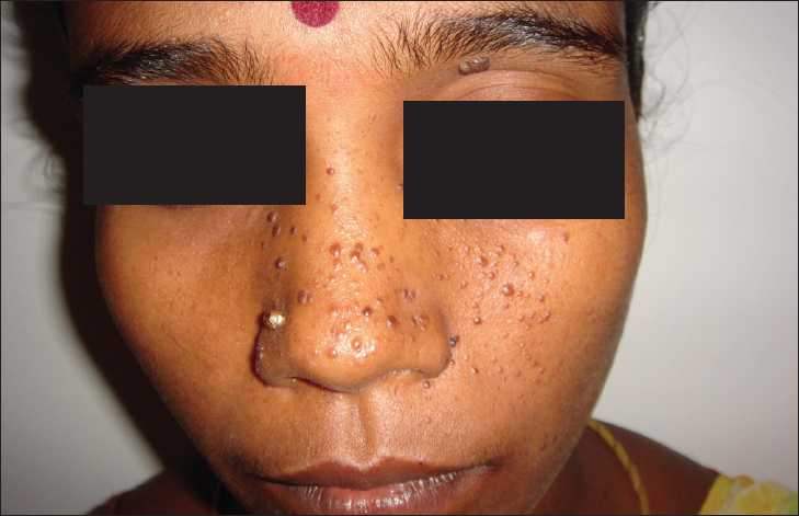
Cherry red papules on the face suggestive of adenoma sebaceum

A reconstructed computerized tomography-abdomen film showing a mass lesion (arrow) in the upper pole of the left kidney with fat attenuation suggestive of renal angiomyolipoma
Tuberous sclerosis is a common neuro-cutaneous syndrome characterized by the triad of adenoma sebaceum, seizures and mental retardation.[1] Adenoma sebaceum is present in most individuals with TS while the incidence of seizures and mental retardation is about 62 and 38% respectively.[2] Additional skin manifestations include ash-leaf spots, shagreen patch and ungual fibromas.[2] TS is a multi-system disorder and is associated with hamartomas and subependymal astrocytoma of the brain, retinal phakoma, cardiac rhabdomyoma and lymphangiomyomatosis of the lung.[1]
Renal lesions include angiomyolipoma, renal cyst and a marginal high incidence of renal cell carcinoma.[1] Pain is the most frequent symptom of renal angiomyolipoma, which may be complicated by life threatening hemorrhage. Renal angiomyolipoma greater than 4 cm should be angiographically studied and treated with partial nephrectomy or renal artery embolization to prevent the dreaded bleeding complication.[1] Adenoma sebaceum can be treated with cryosurgery or laser with good cosmetic results.[34]
Source of Support: Nil
Conflict of Interest: None declared.
References
- Recognizing an index case of tuberous sclerosis. Am Fam Physician. 2000;61:703-8. 710
- [Google Scholar]
- Successful treatment of adenoma sebaceum with the potassium titanyl phosphate laser. Clin Exp Dermatol. 1998;23:201-3.
- [Google Scholar]
- Treatment of adenoma sebaceum with the copper vapor laser. J Am Acad Dermatol. 1995;33:770-4.
- [Google Scholar]


