

Translate this page into:
Comparison of serum creatinine and spot urine interleukin-18 levels following radiocontrast administration
This is an open-access article distributed under the terms of the Creative Commons Attribution-Noncommercial-Share Alike 3.0 Unported, which permits unrestricted use, distribution, and reproduction in any medium, provided the original work is properly cited.
This article was originally published by Medknow Publications & Media Pvt Ltd and was migrated to Scientific Scholar after the change of Publisher.
Abstract
Radiocontrast administration is an important cause of acute renal failure. In this study, compared the plasma creatinine levels with spot urine IL-18 levels following radiocontrast administration. Twenty patients (11 males, 9 females) underwent radiocontrast diagnostic and therapeutic-enhanced examinations. The RIN Mehran risk score was low (≤5). The radiocontrast agents used were 623 mg/mL Iopromid (1.5 mL/kg), and 100 mL of 650 mg/mL meglumine diatrizoate as three-way oral and rectal contrast material for abdominal computed tomography (CT) scans. Serum blood urea nitrogen, creatinine, Na, K, Cl, Ca, P, creatinine clearance, and spot urine IL-18 levels were analyzed before and repeated at 24, 48, and 72 h after radiocontrast administration. Six and 24-h urinary IL-18 levels were measured with a human IL-18 ELISA kit following radiocontrast administration. An increase in plasma creatinine 24 and 48 h following radiocontrast administration was observed compared with precontrast values, but it was not statistically significant (P=0.052 and P=0.285, respectively). A statistically significant increase in IL-18 levels was observed at 6 and 24 h, compared with precontrast values (P=0.048 and P=0.028, respectively). A tendency for postcontrast 24-h urinary IL-18 levels to increase was observed compared with 6 h, but the increase was not statistically significant (P=0.808). Our results show that plasma creatinine starts to increase at 24th hour; however, spot urine IL-18 levels go up at 6th hour following radiocontrast administration implying urine IL-18 to be an earlier parameter for kidney injury.
Keywords
Radiocontrast
serum creatinine
urine IL-18

Introduction
Radiocontrast-induced nephropathy (RIN) can lead to acute renal failure (ARF), which may require dialysis therapy. ARF increases treatment cost due to sepsis, hemorrhage, respiratory failure, and a long hospitalization.[1–3] RIN is an important cause of hospital-acquired ARF and is responsible for 12% of cases.[45] Renal medullary hypoxia and the direct toxic effects of iodinated contrast agents on renal tubules are possible mechanisms responsible for the pathophysiology of RIN.[6] Identified specific risk factors for RIN are current renal insufficiency, diabetes mellitus, and high contrast volume, dehydration, advanced age (>70 years), congestive heart failure (CHF), and nephrotoxic drug use (angiotensin converting enzyme inhibitor and nonsteroidal antiinflammatory drugs).[6] The incidence of RIN with no risk factor is approximately 3–5%.[5] Patients at risk for radiocontrast nephropathy are recommended to use nonionic iso-osmolar or nonionic low osmolar contrast agents.[7] Increases in serum creatinine levels are useful for detecting RIN. In the majority of patients, plasma creatinine levels rise within the first 24–48 h after administering the radiocontrast agent, reach a peak within 3–5 days, and return to normal after 1–3 weeks.[8] Recent studies have reported that urine levels of IL-18 (a pro-inflammatory cytokine), kidney injury molecule-1, and neutrophil gelatinase-associated lipocalin (NGAL) levels are important for early detection of RIN.[910] IL-18 is thought to reveal renal tubular damage due to ischemia.[11] Some reports have shown that IL-18 levels start to increase within 4–6 h and peak at 12 h in patients with acute renal injury.[1213] In this study, we aimed to compare the plasma creatinine levels with spot urine IL-18 levels following radiocontrast administration.
Patients and Methods
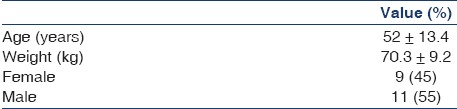
Twenty patients (11 males and 9 females) underwent diagnostic and therapeutic contrast-enhanced examinations at the Department of Internal Medicine from January 2009 to March 2009. The study was approved by the Institute Ethics Committee and written consent was obtained from the selected patients based on a low Mehran risk score (≤5).[14] Demographic characteristics are given in Table 1.
A precontrast-enhanced examination of serum blood urea nitrogen (BUN), creatinine, Na, K, Cl, Ca, P, creatinine clearance was analyzed and they were repeated at 24, 48, and 72 h following contrast administration. Spot urine IL-18 levels were measured before and 6 and 24 h after radiocontrast administration with a human IL-18 ELISA kit (Biosource Invitrogen Human IL-18, California).
Intravenous iopromid (623 mg/mL, 1.5 mL/kg; Ultravist 300), a three-way oral and rectal contrast material for abdominal CT scans, and 650 mg/mL meglumine diatrizoate (Urovist, 100 mL) were used for every patient. Glomerular filtration rate was calculated using the Cockcroft–Gault equation.
1 h before the procedure, 8.4% NaHCO3 plus 5% dextrose (3 mL/kg/h) with 1200 mg/day N-acetylcysteine was given to all patients prophylactically. After radiocontrast agent administration, the same prophylactic treatment was continued (1 mL/kg/h) for 6 h. During this time, hydration and urine output were followed by monitoring the intake and release of fluids. Contrast-enhanced CT was obtained based on the indications.
Patients with no history of kidney disease, plasma creatinine values <1.2 mg/dl, GFR≥60 mL/min, nondiabetic, no urinary infection, and no decompensated heart failure were included. Urinary IL-18 levels were measured with a human IL-18 ELISA kit (Biosource Invitrogen Human IL-18, Carlsbad, CA, USA).
The statistical analysis was performed with the NCSS PASS 2007 and 2008 statistical software (Kaysville, UT, USA). Results are evaluated with 95% confidence intervals. The significance level was 0.05.
Results
Serum creatinine levels increased after radiocontrast administration compared with precontrast levels, although the result was not statistically significant [Table 2]. A slight increase in creatinine levels occurred at 48 h after radiocontrast administration but they fell to precontrast values at 72 h. A slight increase in plasma creatinine levels at 24 and 48 h following radiocontrast administration was observed compared with precontrast values, but it was not statistically significant (P=0.052 and P=0.285, respectively) [Table 2].

Compared with precontrast urine spot IL-18 levels, postcontrast 6 and 24 h urinary levels of IL-18 increased significantly (P=0.048 and P=0.028, respectively; Table 3). A tendency for postcontrast 24-h urinary IL-18 levels to increase was observed compared with 6 h, but the increase was not statistically significant (P=0.808; Table 3).

Plasma creatinine levels and spot urine IL-18 were weakly but positively correlated with those during the precontrast period, although this finding was not statistically significant (r=0.246, P=0.126). Similarly, postcontrast 24-h plasma creatinine levels and spot urine IL-18 levels were weakly but positively correlated, although this result was also not statistically significant (r=0.254, P=0.276).
There was no difference between pre- and postcontrast values of serum blood urea nitrogen (BUN), creatinine, Na, K, Cl, Ca, P, and creatinine clearance.
Discussion
RIN-related high mortality necessitates an early diagnosis and prevention.[15] The RIFLE criteria allow assessment of postcontrast kidney damage.[1617] The most common definition of RIN is plasma creatinine levels of 0.5 mg/dL or higher 72 h after contrast administration or 25% higher than the basal plasma creatinine level.[18] Plasma creatinine levels began to rise within 24 h in 80% of the patients with RIN, peaking at 48–72 h, and returning to baseline after 2 weeks.[4] The first 24 h remains unclear in patients with acute renal injury, but IL-18 levels start to increase within 4–6 h, peaking at 12 h.[1213] Additionally, plasma creatinine is affected by age, body weight, total body volume, gender, race, drug use, muscle mass, and protein intake so researchers are looking for a diagnostic marker for RIN that can be measured easily and is not affected by nonrenal factors.[9]
Most studies related to these parameters include serum and urine cystatin C, serum and urine NGAL, and urine IL-18 in the analysis.[9] As proinflammatory cytokine IL-18 levels increase in urine, tubular inflammation, such as ischemia, reperfusion injury, allograft rejection, cisplatin toxicity, and endotoxemia occur.[1019] Parikh et al., found that 72 patients with acute tubular necrosis and delayed graft reaction have significantly higher IL-18 levels than other kidney diseases (urinary tract infection, chronic renal failure, nephrotic syndrome, or prerenal azotemia).[11] Coca et al. reported that IL-18 generally showed a low sensitivity but high specificity, respectively, for assessing an acute kidney injury diagnosis and risk classification.[9]
In our study, we examined spot urinary IL-18 levels in comparison with plasma creatinine levels. A weak positive correlation was found between precontrast creatinine and urine IL-18 levels, although it was not statistically significant. Furthermore, we also found a weak positive correlation between postcontrast 24 h creatinine and urine IL-18 levels, although this was not statistically significant either. A slight increase in plasma creatinine levels at 24 h and 48 h following radiocontrast administration was observed compared with precontrast values, but it was not statistically significant which was regressed to precontrast values at 72 h. A statistically significant increase in the level of spot urinary IL-18 levels at 6 and 24 h postcontrast was observed, compared with precontrast spot urine IL-18 levels and difference between 6th and 24th hour levels were not statistically significant.
One of the limitations of this study is that there are other early biomarkers of acute kidney injury such as NGAL[20] and KIM-1.[21] Another limitation is that urine IL-18 measurement was indexed to serum creatinine instead of urine creatinine since serum creatinine is considered to be a better and commonly used marker in the diagnosis of acute kidney injury.
In conclusion, spot urine IL-18 levels at sixth hour following radiocontrast administration suggesting that it may be an earlier parameter for identifying kidney injury.
Source of Support: Nil
Conflict of Interest: None declared.
References
- Pathophysiology and Etiology of Acute Renal Failure. In: Feehally J, Floege J, Johnson JR, eds. Comprehensive Clinical Nephrology (3rd ed). Philadelphia: Mosby Elsevier; 2007. p. :755-70.
- [Google Scholar]
- Renal failure after major angiography can be avoided with hydration. AJR Am J Roentgenol. 1981;136:859-61.
- [Google Scholar]
- Hospital-acquired renal insufficiency: A prospective study. Am J Med. 1983;74:243-8.
- [Google Scholar]
- Nephropathy induced by contrast media: Pathogenesis, risk factors and preventive strategies. CMAJ. 2005;172:1461-71.
- [Google Scholar]
- Incidence and prognostic importance of acute renal failure after percutaneous coronary intervention. Circulation. 2002;105:2259-64.
- [Google Scholar]
- Contrast media nephropathy –how to diagnose and how to prevent? Neph Dial Transpl. 2007;22:1812-5.
- [Google Scholar]
- Biomarkers for the diagnosis and risk stratification of acute kidney injury: A systemic review. Kidney Int. 2008;73:1008-16.
- [Google Scholar]
- Urinary biomarkers for acute kidney injury: Perspectives on translation. Clin J Am Soc Nephrol. 2008;3:481-90.
- [Google Scholar]
- Urinary interleukin-18 is a marker of human acute tubular necrosis. Am J Kidney Dis. 2004;43:405-14.
- [Google Scholar]
- İmprovements in the diagnosis of acute kidney injury. Saudi J Kidney Dis Transpl. 2008;19:537-44.
- [Google Scholar]
- IL-18 contributes to renal damage after ischemia-reperfusion. J Am Soc Nephrol. 2008;19:2331-41.
- [Google Scholar]
- A simple risk score for prediction of contrast-induced nephropathy after percutaneous coronary intervention: Development and initial validation. J Am Coll Cardiol. 2004;44:1393-9.
- [Google Scholar]
- Canadian Association of Radiologists: Consensus quidelines for the prevention of contrast –induced nephropathy. Can Assoc Radiol J. 2007;58:79-87.
- [Google Scholar]
- The clinical and renal consequences of contrast-induced nephropathy. Nephrol Dial Transplant. 2006;21:i2-10.
- [Google Scholar]
- Defining acute renal failure: RIFLE and beyond. Clin J Am Soc Nephrol. 2006;1:1314-9.
- [Google Scholar]
- Contrast-media-induced nephrotoxicity: A consensus report. Contrast Media Safety Committee, European Society of Urogenital Radiology (ESUR) Eur Radiol. 1999;9:1602-13.
- [Google Scholar]
- Serum neutrophil gelatinase-associated lipocalin (NGAL) in predicting worsening renal function in acute decompensated heart failure. J Card Fail. 2010;16:49-54.
- [Google Scholar]
- Kidney injury molecule-1 (KIM-1): A urinary biomarker and much more. Nephrol Dial Transplant. 2009;24:3265-8.
- [Google Scholar]


