

Translate this page into:
Percutaneous stent-in-stent placement for renal artery stenosis of a solitary functioning kidney
This is an open-access article distributed under the terms of the Creative Commons Attribution-Noncommercial-Share Alike 3.0 Unported, which permits unrestricted use, distribution, and reproduction in any medium, provided the original work is properly cited.
This article was originally published by Medknow Publications & Media Pvt Ltd and was migrated to Scientific Scholar after the change of Publisher.
Abstract
Renal artery stenosis is an important cause of secondary hypertension in young, which can be treated effectively with endovascular interventions including stenting. In-stent thrombosis is an infrequent complication and necessitates the need to follow-up these patients with renal artery stents. We present here a young individual who presented with in-stent thrombosis in a solitary functioning kidney and was successfully treated with stent-in-stent placement.
Keywords
Renal angiogram
renovascular hypertension
stent thrombosis

Introduction
Renal artery stenosis is one among a wide variety of uncommon, but potentially treatable causes of secondary hypertension.[1] Atherosclerotic renal artery disease is the most common cause of renal artery stenosis, accounting for 80% of renal arterial lesions. Other causes such as fibromuscular dysplasia, aortic dissection, cholesterol emboli, renal arterial trauma, arteriovenous malformation, polyarteritis nodosa, and Takayasu arteritis form the remaining 20% of renal arterial lesions.
Endovascular renal angioplasty and stenting is the current gold standard treatment for atherosclerotic and fibrous arterial lesions. Re-stenosis is a less recognized long-term complication accounting for about 16% of cases (range: 0-39%).[2] Those patients who undergo renal artery stenting, especially with a single functioning kidney, need periodic evaluation of flow across the stented artery.
Case Report
A 37 year old male presented with a history of hypertension for the last 3 years. He was not a smoker and had no family history of hypertension. The blood pressure was 220/120 mm of Hg despite being on nifedipine 20 mg thrice daily, prazosin 5 mg twice daily and clonidine 0.1 mg 3 times daily. He was found to have severe renal insufficiency (serum creatinine: 8.9 mg/dl), hypokalemia (serum potassium 2.9 meq/l), bland urine sediment, contracted left kidney (6.3 cm × 3.4 cm) and normal sized right kidney (9.4 cm × 4.0 cm) with increased echotexture. The cardiac evaluation showed concentric left ventricular hypertrophy with good LV function (EF: 71%) and mild diastolic dysfunction. Color Doppler showed high resistive indices (RI > 1.0) and peak systolic velocity (>180 cm/sec) in both main renal arteries suggesting ostial stenosis. Renal arteriogram showed hemodynamically significant ostial stenosis on both sides. Renal angioplasty and stenting was carried out on the right side with balloon expandable chromium cobalt stent. He received 5000 units of unfractionated heparin in the post procedure period and was started on anti-platelet agents. His serum creatinine decerased to 1.5 mg/dl over the next 4 weeks. The blood pressures were within the normal range on 40 mg of nifedipine daily. He continued to use anti-platelet agents (aspirin 150 mg and clopidogrel 75 mg daily).
He presented with flash pulmonary edema after 1½ years of initial stent placement. When his follow-up visits with the local physician were reviewed it revealed a slowly rising blood pressure levels for which an angiotensin converting enzyme inhibitor (ramipril 5 mg) was added in the last 2 weeks. Re-evaluation showed severe renal insufficiency (serum creatinine 10.2 mg/dl). Color Doppler showed no flow across the stent. Renal arteriogram showed near total stent occlusion with no collateral flow into the kidney [Figure 1].

- Stent - in - stent with well perfused kidney
Ramipril was discontinued and he was given two sessions of hemodialysis. A percutaneous balloon dilatation and stent-in-stent (Medtronic racer: 5.0 mm × 18 mm, cobalt chromium) was placed [Figures 2 and 3]. On the 2nd day of stent placement his urine out put improved and serum creatinine started to decline and reached a nadir of 1.5 mg/dl on the 7th day.
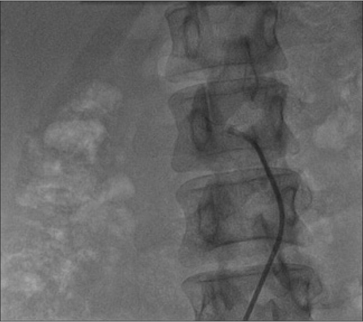
- Right renal artery stent with stent thrombosis

- Right renal artery with balloon across the thrombotic segment in the existing stent
Discussion
Early revascularization of a solitary functioning kidney not only improves blood pressure, but also the kidney function. Acute and long-term complications of the endovascular interventions vary between institutions depending on the available expertise. Although complications are not common, they can be catastrophic, including a theroembolic disease and aortic dissection.[1]
The potential benefits of early intervention can be seen either in the form of blood pressure control or salvage of renal function in ischemic nephropathy. Large observational studies indicate either cure (12%) or improvement (73%) in hypertension rates.[34] These studies also showed that either there is an improvement (30%) or stabilization (42%) of renal function after early intervention.[34]
Our patient responded well to initial stent placement before the onset of ischemic nephropathy with prompt reduction of blood pressure and improvement of renal function, which were probably as a result of critical stenosis.
Re stenosis is an important and considerable draw back in the long-term. A meta-analysis by Leertouwer et al.,[5] showed the incidence of re stenosis as 16% over a follow-up period of 20 months.
There are no clear guidelines for follow-up of stented patients. The appropriate treatment strategy for in-stent stenosis has not yet been defined. Evidence provided by Zeller T et al.,[6] indicates that in-stent stenosis should be treated with stent-in-stent placement favoring long-term patency rather than angioplasty alone. The likelihood of re stenosis depends on individual factors such as vessel diameter, compliance with anti-platelet agents, bilateral renal artery stenosis etc.
Elevated blood pressures in a hitherto controlled patient and worsening of renal function following the introduction of ACEi in our patient who has a solitary functioning kidney previously stented points toward the diagnosis of in-stent stenosis. Following the establishment of diagnosis by renal arteriogram, our patient showed good clinical response in the form of improvement of blood pressure and renal function after balloon angioplasty and stent-in-stent placement. This report shows the importance of follow-up of renal stents with duplex ultrasound for evidence of restensosis.
Source of Support: Nil
Conflict of Interest: None declared.
References
- Controversies in renal artery stenosis: A review by the American Society of Nephrology Advisory Group on Hypertension. Am J Nephrol. 2007;27:212-20.
- [Google Scholar]
- Management of reno vascular disease: A review of renal artery stenting in ten studies. QJM. 1999;92:159-67.
- [Google Scholar]
- Stents in the treatment of renal artery stenosis: Long-term follow-up. J Endovasc Surg. 1999;6:42-51.
- [Google Scholar]
- Renal vascular disease: Medical management, angioplasty, and stenting. Semin Nephrol. 2000;20:474-88.
- [Google Scholar]
- Stent placement for renal arterial stenosis: Where do we stand. A meta-analysis? Radiology. 2000;216:78-85.
- [Google Scholar]
- Treatment of in stent re stenosis following stent-supported renal artery angioplasty. Catheter Cardiovasc Interv. 2007;70:454-9.
- [Google Scholar]


