

Translate this page into:
Unusual finding of cellular glomeruli infiltrated with lymphocytes in end stage renal disease due to obstructive nephropathy
This is an open-access article distributed under the terms of the Creative Commons Attribution-Noncommercial-Share Alike 3.0 Unported, which permits unrestricted use, distribution, and reproduction in any medium, provided the original work is properly cited.
This article was originally published by Medknow Publications & Media Pvt Ltd and was migrated to Scientific Scholar after the change of Publisher.
Sir,
A 15-year-old female presented with complaints of right flank pain since 2 months. Per abdomen examination revealed tenderness in right lumbar region on bimanual palpation. Ultrasonography and computerized tomography showed a mildly hydronephrotic kidney on right side with a calculus in the pelvis of kidney. Diethylene triamine pentaacetic acid renal scan showed a non-functioning right kidney with glomerular filtration rate of 1.8 ml/min and a 2.7% split function. It also showed an optimally functioning left kidney. Patient underwent a laparoscopic right nephrectomy. Grossly kidney measured 4.5 cm × 3 cm × 1.7 cm. On cutting cortex was atrophic with dilatation of pelvicalyceal system and a calculus was found in the pelvis measuring 1 cm × 0.5 cm × 0.5 cm. Microscopic sections of kidney showed cortical scarring, atrophy, interstitial chronic inflammatory infiltrate and focal tubular thyroidization. Glomeruli were cellular and on higher magnification showed infiltration with lymphocytes which on immunohistochemistry were seen to be mixture of B and T lymphocytes [Figure 1].
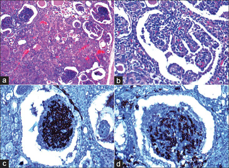
- (a) The cellular glomeruli with chronic interstitial inflammatory infiltrate and thyroidized tubules (H and E, ×100). (b) The cellular glomerulus with lymphocytic infiltration (H and E, ×400). (c and d) Immunoreactivity for CD20 and CD3 (×400)
There are two possible explanations for the above finding of cellular glomeruli with an infiltrate of lymphocytes in them.
One hypothesis states that there is a glomerulotubular feedback loop that results in progression of chronic kidney disease.[1] It is postulated that the protein in the ultra-filtrate gets degraded by tubular epithelial cells and possibly directly processed by dendritic cells and is presented to the T lymphocytes.[23] These proteins consist of multiple self-antigens and other proteins and peptides such as fragments of glomerular basement membrane and other glomerular antigens. In the normal state dendritic cell uptake of antigen and cross presentation will lead to apoptosis of CD8 + cells resulting in induction of tolerance. In the presence of injured cells, the process of cross presentation to CD8 + cells results in active immunity, secondary to so called danger signals or damage associated molecular patterns (DAMPs). The presence of DAMPs results in CD4 + helper and CD8 + cytotoxic lymphocyte activation and initiation of tissue injury and autoimmunity, thus amplifying original damage and leading to progression of organ damage.[1]
In contrsst, Pascal et al. suggested that unilateral obstruction produced a tubular injury that led to recognition of some unidentified antigen by the immune system. This resulted in an antibody response and formation of antigen-antibody complexes which were deposited in the glomeruli and led to glomerulopathy.[4] A similar sequence of events is possibly the explanation for the glomerular lesions found in our case.
References
- T cells and dendritic cells in glomerular disease: The new glomerulotubular feedback loop. Kidney Int. 2010;77:393-9.
- [Google Scholar]
- Proteasomal processing of albumin by renal dendritic cells generates antigenic peptides. J Am Soc Nephrol. 2009;20:123-30.
- [Google Scholar]
- Kidney dendritic cell activation is required for progression of renal disease in a mouse model of glomerular injury. J Clin Invest. 2009;119:1286-97.
- [Google Scholar]
- Demonstration of an antibody to tubular epithelium in glomerulonephritis associated with obstructive uropathy. Am J Med. 1980;69:944-8.
- [Google Scholar]


