

Translate this page into:
Bardet-Biedl syndrome presenting with steroid sensitive nephrotic syndrome
This is an open access article distributed under the terms of the Creative Commons Attribution-NonCommercial-ShareAlike 3.0 License, which allows others to remix, tweak, and build upon the work non-commercially, as long as the author is credited and the new creations are licensed under the identical terms.
This article was originally published by Medknow Publications & Media Pvt Ltd and was migrated to Scientific Scholar after the change of Publisher.
Abstract
Bardet-Biedl syndrome (BBS) is a rare autosomal recessive disorder characterized by postaxial polydactyly, retinitis pigmentosa, central obesity, mental retardation, hypogonadism, and renal involvement. Renal involvement in various forms has been seen in BBS. Cases with nephrotic range proteinuria not responding to steroid have been described in this syndrome. Here we report a case of BBS who presented with nephrotic range proteinuria. The biopsy findings were suggestive of minimal change disease. The child responded well to steroid therapy and remains in remission.
Keywords
Bardet-Biedl syndrome
minimal change nephrotic syndrome
renal anomalies

Introduction
Bardet-Biedl syndrome (BBS) is a rare autosomal recessively inherited disease with prevalence rates of 1:140,000–1:160,000. Higher incidence have been found in isolated populations of Newfoundland and Kuwait (1:1700).[1] This syndrome is characterized by cardinal features, namely central obesity, mental retardation, postaxial polydactyly, pigmentary retinopathy, hypogenitalism and renal involvement.[2] Renal abnormalities include renal dysplasia, fetal lobulation, cystic tubular disease, glomerular disease (most commonly focal segmental glomerulosclerosis). Here we report a rare case of BBS with steroid sensitive, probably minimal change nephrotic syndrome.
Case Report
A 10-year-old male, born of non-consanguineous marriage presented with generalized edema. The parent also complained of short stature, obesity, poor school performance, poor night vision and passing urine from under surface of penis and poor genital development. He had delayed developmental milestones, started walking at the age of 2½ years and spoke bi-syllable word at 3 years of age. There was no history of similar illness in the family. On physical examination, weight was 45 kg (between 90 and 97th centile for age) and height 121 cm (<3rd centile for age) with body mass index of 30.73. The blood pressure was 104/64 mm of Hg. There was brachycephaly, large ears, mild retrognathia, high-arched palate, short and broad hands, post-axial polydactyly in upper limb and left lower limb, micropenis (<2.5 cm) and small testis, hypospadias [Figure 1], central obesity, and anasarca. Respiratory, cardiovascular and abdominal examinations were normal. Fundoscopy revealed retinitis pigmentosa. His intelligence quotient, determined by Stanford Binet method (Indian adaptation by S. K. Kulshrestha), was 70. All these findings were suggestive of BBS.
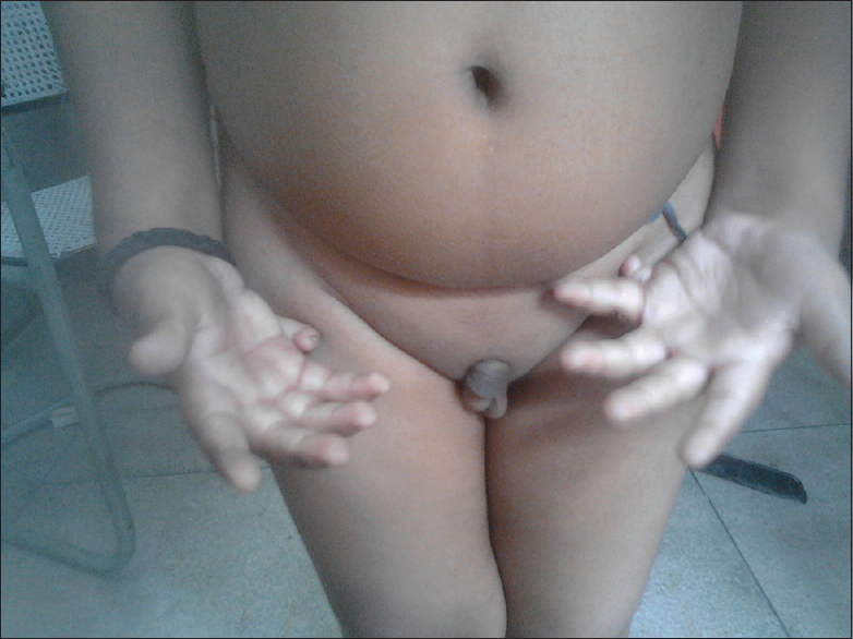
- Polydactyly, micropenis, small scrotum, hypospadias and distended abdomen
His complete blood count was normal. Urinalysis showed nephrotic range proteinuria (3+ on dipstick test), leucocytes 1–2/high power field, and no erythrocytes, with spot urine protein-creatinine ratio of 3.5. The blood urea was 26 mg/dl and serum creatinine was 0.5 mg/dl. The glomerular filtration rate was 99.2 ml/min/1.73 m2. His serum cholesterol was 275 mg/dl. His serum albumin was 2.4 g/dl. Hormonal evaluation showed serum testosterone level of 0.06 ng/dl (normal range = 2–165 ng/dl). His serum electrolytes and random blood sugar were within normal limits. Ultrasonography of whole abdomen showed that both kidneys were normal in size and location with intact corticomedullary differentiations, and bilateral increased echogenicity. His maturating cystourethrography and intravenous pyelography were normal. Echocardiography revealed no abnormalities. In view of significant association renal abnormalities in BBS, a decision to perform renal biopsy was made at the onset of disease itself. Renal biopsy consisted of seven glomeruli, and all appearing essentially unremarkable. The glomerular capillary wall did not show significant thickening. No endocapillary proliferation or neutrophil exudation or segmental glomerular sclerosis was noted [Figure 2]. Tubules exhibited intracytoplasmic vacuolation. Interstitium showed mild inflammation. No tubular atrophy or interstitial fibrosis was seen. The renal biopsy findings were suggestive of minimal change disease. The minimal change nature of the glomerular pathology could not be confirmed as the biopsy specimen contained only seven glomeruli and electron microscopy was not carried out. Immunofluorescence staining for IgA, IgG, IgM, C3, and C1q was negative.

- Hematoxylin and eosin stained biopsy specimen of glomeruli
He was treated with prednisolone and responded remarkably. The patient is in regular follow up for last 8 months and there is no relapse till date.
Discussion
Renal abnormalities, both structural and functional, are common in BBS. Some authors considered it as sixth cardinal feature.[3] Abnormal renal function leading to end stage renal disease is the most common cause of death in these patients.[4] The known renal anomalies include dysplasia, unilateral renal agenesis, cystic tubular disease, fetal renal ectopia, vesicouretric reflux, calyceal clubbing, and blunting. Tubular functional defect results in abnormal urine concentrating ability leading to nephrogenic diabetes insipidus and type 1 renal tubular acidosis.[56] Renal histology has revealed mesangial proliferative glomerulosclerosis, increase in mesangial matrix, and ultrastructural change in glomerular basement membrane and chronic interstitial nephritis.[7] Very rarely, a case of BBS with structural renal anomaly (calyceal clubbing) and nephrotic range proteinuria has been reported.[8] The present case was not associated with any structural anomaly.
The renal biopsy of this case yielded only seven glomeruli, and electron microscopy was not performed. In view of the same, focal segmental glomerulosclerosis cannot be excluded with certainty. That is why, in the present case, we found the term “steroid-sensitive nephrotic syndrome” more preferable to that of “minimal change nephrotic syndrome.”
The presence of steroid-sensitive nephrotic syndrome in a case of BBS may be a feature of the syndrome or may just be an associated finding. BBS can result from mutations in 14 genes (BBS genes). These genes are known to be associated with cell structures called cilia and basal bodies. That is why BBS is now regarded as one of the ciliopathies.[9] The present case report suggests that there seems to be an association between ciliopathy and podocytopathy (presence of nephrotic range proteinuria); more research is needed to explore the exact association. BBS1 gene accounts for one quarter of cases of BBS. In a recent genetic study, researchers found that patients with mutation in BBS6, BBS10, and BBS12 genes had more severe renal disease.[10]
The present case is unique also in the way that the nephrotic syndrome was the presenting feature in this case. On thorough evaluation, BBS was found to be the primary entity. To the best of our knowledge, no case of BBS with documented steroid-sensitive nephrotic syndrome has been reported from India.
Source of Support: Nil
Conflict of Interest: None declared.
References
- High incidence of Bardet Biedl syndrome among the Bedouin. Clin Genet. 1989;36:463-4.
- [Google Scholar]
- The cardinal manifestations of Bardet-Biedl syndrome, a form of Laurence-Moon-Biedl syndrome. N Engl J Med. 1989;321:1002-9.
- [Google Scholar]
- Bardet-Biedl syndrome with end stage kidney disease: A case report and review of literature. Indian J Nephrol. 2007;17:10-3.
- [Google Scholar]
- The spectrum of renal disease in Laurence-Moon-Biedl syndrome. N Engl J Med. 1988;319:615-8.
- [Google Scholar]
- End stage renal disease, differential diagnosis, a rare genetic disorder: Bardet-biedl syndrome: Case report and review. Indian J Clin Biochem. 2011;26:214-6.
- [Google Scholar]
- Ultrastructural changes in the glomerular basement membrane of patients with Laurence-Moon-Biedl-Bardet syndrome. Clin Nephrol. 1981;16:283-8.
- [Google Scholar]
- Glomerulonephropathy of Laurence-Moon-Biedl syndrome. Postgrad Med J. 1988;64:621-5.
- [Google Scholar]
- Basal body dysfunction is a likely cause of pleiotropic Bardet-Biedl syndrome. Nature. 2003;425:628-33.
- [Google Scholar]
- Bardet-Biedl syndrome: A study of the renal and cardiovascular phenotypes in a French cohort. Clin J Am Soc Nephrol. 2011;6:22-9.
- [Google Scholar]


