

Translate this page into:
Renal infarct following varicella infection
This is an open access article distributed under the terms of the Creative Commons Attribution-NonCommercial-ShareAlike 3.0 License, which allows others to remix, tweak, and build upon the work non-commercially, as long as the author is credited and the new creations are licensed under the identical terms.
This article was originally published by Medknow Publications & Media Pvt Ltd and was migrated to Scientific Scholar after the change of Publisher.
Abstract
Renal infarction usually occurs against a background of heart disease or a thromboembolic tendency and rarely is associated with infections. Here we present a case of a young boy who reported with painless gross hematuria following primary Varicella infection and was found to have an isolated renal infarct.
Keywords
Hematuria
renal infarct
varicella infection

Introduction
Acute renal infarction is rarely detected in clinical practice. The diagnosis is often missed or delayed due to its non-specific clinical presentation.[1] We are reporting the first case of acute renal infarction after varicella infection which presented as painless hematuria.
Case Report
A 20 year-old boy presented with fever for 2 days with centripetally distributed vesicular lesion that started from chest and progressed to extremities. Fever subsided over 3 days and the lesions started to scab after 7 days. A week later he complained of left sided flank pain, which continued for 48 h followed by development of gross painless hematuria. Hematuria persisted for 72 h without any accompanying lithuria, graveluria or pain. The patient denied consumption of nonsteriodal anti-inflammatory agents or any antibiotic. Investigations revealed hemoglobin (Hb) 15.5 gm/dL, total leukocyte count 4600/mm3, Platelets 2.3 lacs/mm3, erythrocyte sedimentation rate 18 mm fall in 1st hour, Urine analysis revealed 2+ albumin with numerous red blood cells (RBC)/hpf; however there were no dysmorphic RBC's, blood urea 38mg/dL, serum creatinine 0.9 mg/dL, prothrombin time (Test 13.0 sec/control 13.0 sec/international normalized ratio 1.0). Sonography of kidney was normal. Renal Doppler revealed normal flow in right kidney; however there was no flow in left segmental renal artery supplying lower pole. Computed tomography (CT) Renal angiography revealed a wedge shape defect in left kidney suggestive of infarct with no flow in segmental renal artery [Figure 1]. Vasculitic workup was negative for any underlying autoimmune disease (C – reactive protein <0.6 mg/dl, rheumatoid factor 11.5 U/ml, normal complement, negative anti nuclear antibody/double stranded DNA and anti neutrophil cytoplasmic antibodies). Hemoglobin electrophoresis ruled out possibility of sickle cell anemia. Serology for varicella confirmed recent infection with IgM varicella with a value of 17.54 U/mL (normal <8 U/mL). Limited workup for procoagulant state did not found anything significant (negative rlupus anticoagulant test by dilute russel viper venom time and negative IgM/IgG anti phospholipid antibodie). The diagnosis of varicella vasculopathy leading to renal infarction was considered in view of serological diagnosis of recent varicella infection and temporal appearance of symptoms. This patient was managed conservatively. He has been on regular follow-ups and has preserved renal function with no fresh hematuria.
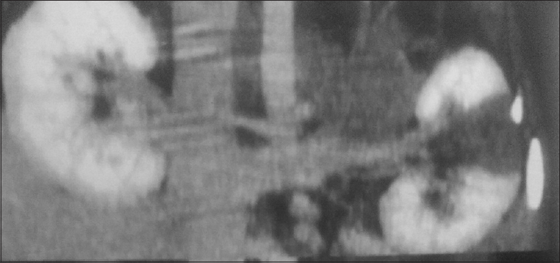
- Computed tomography renal angiography showing wedge-shaped infarct in renal parenchyma
Discussion
The diagnosis of acute renal infarction is often missed, mainly due to the entity's non-specific clinical presentation and lack of physician awareness. Renal infarction may present as subtle symptoms such as nausea, vomiting, fever, hypertension or overt symptoms abdominal pain or hematuria; however it may remain asymptomatic. We present first documented a case of varicella zoster-associated vasculopathy, presenting as isolated renal infarction.
Most cases of renal infarction documented in literature have been usually associated with heart diseases, underlying hypercoagulable state or collagen vascular disease. Huang et al. in their case series reported that the most common cause for renal infarction was underlying cardiac condition most likely atrial fibrillation and diagnosis of renal infarction was confirmed on CT renal angiography.[2] In our case also although obstructed flow on segmental renal artery was picked by Doppler, confirmation of renal infarct was made on CT angiography. Varicella infection leading to vasculitic complications is rare. Though varicella vasculopathy is an infection related vasculitis, it usually affect medium and large vessels, mostly cerebral vasculature. It commonly presents with acute stroke, ataxia or blindness; however involvement of peripheral vessels is infrequent.[3] The diagnosis of varicella vasculopathy usually rest on the temporal occurrence of the symptoms following varicella infection.[4]
The exact mechanism leading to vasculitis has not been elucidated. It is believed that it results from damage to the vessel wall media by direct viral invasion, immune-complex reactions, or a combination of both.[4] This is the first such case in literature where primary varicella infection was associated with isolated renal vasculitis. In tropical countries such as ours where infection related vasculitic complications are common, patients presenting with hematuria following episode fever and rash possibility of varicella associated vasculopathy should also be considered.
Financial support and sponsorship
Nil.
Conflicts of interest
There are no conflicts of interest.
References
- Clinical characteristics of renal infarction in an Asian population. Ann Acad Med Singapore. 2008;5:416-20.
- [Google Scholar]
- Varicella zoster virus vasculopathies: diverse clinical manifestations, laboratory features, pathogenesis, and treatment. Lancet Neurol Aug. 2009;8:731.
- [Google Scholar]
- The epidemiology of varicella in staff and students of a hospital in torpics. Int J Epidemiol. 1984;13:502-5.
- [Google Scholar]


