

Translate this page into:
Urinary Neutrophil Gelatinase-Associated Lipocalin and Urinary Soluble CXCL16 as Biomarkers of Activity in Pediatric Lupus Nephritis
This is an open access journal, and articles are distributed under the terms of the Creative Commons Attribution-NonCommercial-ShareAlike 4.0 License, which allows others to remix, tweak, and build upon the work non-commercially, as long as appropriate credit is given and the new creations are licensed under the identical terms.
This article was originally published by Medknow Publications & Media Pvt Ltd and was migrated to Scientific Scholar after the change of Publisher.
Abstract
One of the challenges of treating patients with lupus nephritis (LN) is to assess disease activity. The aim of this study was to measure the urinary neutrophil gelatinase-associated lipocalin (uNGAL) and urinary soluble chemokine (C-X-C motif) ligand 16 (CXCL16) levels in children and adolescents with systemic lupus erythematosus (SLE) and investigate whether they are elevated in active LN. This study was conducted on 80 patients diagnosed as SLE by the Systemic Lupus International Collaborating Clinics criteria and 60 apparently healthy individuals as controls. Global and renal disease activities were evaluated by Systemic Lupus Erythematosus Disease Activity Index (SLEDAI) and renal SLEDAI, respectively. uNGAL and urinary CXCL16 were measured for all participants by ELISA. Renal biopsy was done for all cases at initial diagnosis and was graded using ISN/RPS classification. uNGAL and CXCL16 were higher in patients than in the controls (8.9 ± 3.56 ng/dl and 1067 ± 367 ug/L vs. 2.26 ± 1.95 ng/dl and 471 ± 106 ug/L, respectively). uNGAL had higher sensitivity and specificity than urinary CXCL16 as predictor of LN (95% and 90% vs. 85% and 80%, respectively). There was significant positive correlations between uNGAL levels, 24-h urinary proteins (r = 0.732, P = 0.001), and SLEDAI (r = 0.359, P = 0.001). There was also significant positive correlations between urinary CXCL16 levels, 24-h urinary proteins (r = 0.47, P = 0.001), and SLEDAI (r = 0.17, P = 0.001). uNGAL and CXCL16 were reliable indicators of the activity of LN.
Keywords
Activity
CXCL16
nephritis
systemic lupus erythematosus
urinary neutrophil gelatinase-associated lipocalin

Introduction
Lupus nephritis (LN) is considered the most serious manifestation of systemic lupus erythematosus (SLE) and can be present in 60% of SLE patients.[1] Kidney biopsy is usually done in patients with abnormal urinary findings, e.g., sediment, proteinuria, or elevated serum creatinine. These markers do not always correlate with histopathological diagnosis. Kidney involvement is known to precede the appearance of proteinuria, elevation of serum creatinine, or abnormal urine sediment leading to a delay in the management of LN, its flares and assessment of response to therapy, thus leading to more morbidity and mortality.[2] Reliable biomarkers may help in the evaluation of disease activity, identify patients at risk for organ damage or recurrent flares, and facilitate early and accurate evaluation of responses to treatment.[2]
Neutrophil gelatinase-associated lipocalin (NGAL)[3] was identified as one of the earliest and most robustly induced genes and proteins in the kidney after ischemic or nephrotoxic injury and was easily detected in the urine and blood soon after acute kidney injury.[34] Urinary NGAL (uNGAL) was proven as an excellent biomarker of concurrent LN activity. In spite of the fact that NGAL can go up in any renal injury and may not be specific for SLE nephritis, this promising biological marker can point to acute kidney injury in patients with SLE.[4]
Serum soluble chemokine (C-X-C motif) ligand 16 (CXCL16) was recently reported to be selectively upregulated in a wide range of tissues in response to damage, especially in kidney[5] CXCL16 has been documented to be increased within the kidneys, sera, and urine of SLE patients and seems to correlate with disease activity, although their clinical utility in predicting disease activity in LN remains unclear.[5]
We aimed in this research to measure uNGAL and CXCL16 levels in children and adolescents with SLE and investigate whether they were elevated in active SLE patients and to verify their role in the diagnosis of LN.
Methods
Sample size
The sample size was calculated taking into consideration a significance level of 95%, power 80%, and effect size 30% resulting in 86 children to be included. A sample frame which consists of the files of all unit attendants who fulfill the selection criteria was constructed from which the target population was chosen randomly, where only 80 children participated in our study (Six patients refused to participate).
The cross-sectional, observational study was conducted after approval from the Ethical Committee of the Faculty of Medicine, Tanta University, and informed written parental consents from 80 patients diagnosed as SLE was taken. They were selected from the inpatient department and outpatient clinic of the Pediatric Nephrology Unit of the Pediatric Department, Tanta University Hospital, between July 2016 and July 2017.
Inclusion criteria
All patients previously or currently fulfilled criteria for the diagnosis of SLE.[6]
A total of 60 apparently healthy age and sex matched individuals as healthy controls were also chosen. All patients and controls were subjected to:
-
Full history taking
-
Thorough clinical examination.
Global disease activity was evaluated according to the Systemic Lupus Erythematosus Disease Activity Index (SLEDAI) score. Renal disease activity was evaluated according to the renal SLEDAI (rSLEDAI).[7]
Urinary neutrophil gelatinase-associated lipocalin and urinary CXCL16
They were measured by the enzyme-linked immunosorbent assay using Human ELISA Kit (Epitope Diagnostics, Inc., San Diego, USA) as per the manufacturer's protocol. (Range of standard was 0.31–4.21 nanogram/deciliter).
Sampling
All urine samples were diluted 1:1 for the ELISA, and the concentrations of the respective molecules were ascertained from the standard curves constructed using manufacturer-supplied standards. In the Biovendor Human NGAL ELISA, standards, quality controls, and samples were incubated in microplate wells with polyclonal antihuman NGAL antibody. After 1 h incubation and washing, biotin-labeled polyclonal antihuman NGAL antibody was added and incubated with captured NGAL for 1 h. After another washing, streptavidin-horseradish peroxidase conjugate was added. After 30 min incubation and the last washing step, the remaining conjugate was allowed to react with the substrate solution (TMB). The reaction was stopped by addition of acidic solution and absorbance of the resulting yellow product was measured spectrophotometrically at 450 nm.
Urinary CXCL16 concentrations were quantified using a double-ligand ELISA methodology, according to the manufacturer's instructions (chemokine domain, R&D System, Minneapolis, MN, USA).
Renal biopsy
The kidney biopsies were done for all cases at initial diagnosis to determine the severity of LN.[8]
Statistical analysis
Patients’ data were tabulated and processed using SPSS V16. (Methodology, Methods and technigues, 2nd edition. New Delhi, India)[9] software statistical computer package. The difference between parametric data of groups analyzed through Student's t-test and ANOVA test. Chi-square test was used for comparison of frequencies. Spearman's correlation coefficient (r) was used to assess the degree of association between 2 continuous variables. To assess the diagnostic value, we calculated the following indices: cutoff value, accuracy, sensitivity, specificity, and positive predictive value, that is, the proportions of the positive results that are true positive and negative predictive value, that is, the proportions of the negative results that are true negative. A value of (P < 0.05) considered statistically significant.[9]
Results
The demographical and laboratory data of the studied groups are summarized in Table 1. The total number of the SLE patients was 80:70 females (87.50%) and 10 males, F:M ratio (7:1). Their age had a mean of 12.07 ± 3.35 years. A total of 60 apparently healthy individuals were carefully chosen, matched for age and sex with the SLE patients. They were 52 female (86.67%) and 8 male, F:M ratio (6.5:1). Their mean age was 12.32 ± 3.23 years, with no significant differences between the patients and controls. SLEDAI score has been used in our study as a predictor of disease activity. The median of global SLEDAI score was 26.8, rSLEDAI was 10.3, and extra-rSLEDAI was 16.8. Renal biopsies at initial diagnosis evaluated according to the ISN LN grading system as shown in Table 2, ISN Classes I (50%) and III (35%) were the most common findings.
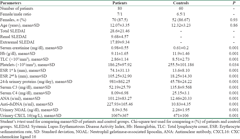

The mean concentrations of uNGAL (8.9 ± 3.56 ng/dl) and urinary CXCL16 (1067 ± 367 ug/L) in SLE patients were significantly higher than the controls (P < 0.05). Table 3 compared between the mean levels of uNGAL and urinary CXCL16 in SLE patients with renal affliction according to renal biopsy results. There was significant difference between mean levels of uNGAL and urinary CXCL16 in SLE patients with the renal involvement of different classes according to renal biopsy results (P < 0.05). Table 4 and Figure 1 summarized validity of uNGAL and urinary CXCL16 in prediction of LN. uNGAL had higher sensitivity and specificity than urinary CXCL16 in early prediction of LN.

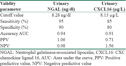

- Receiver operating characteristic curves of urinary neutrophil gelatinase-associated lipocalin and CXCL16 for prediction of activity of lupus nephritis
Table 5 summerized correlations between uNGAL, urinary CXCL16, urinary protein/24-h collected urine, and SLEDAI. There was a significant positive correlation between uNGAL levels and 24-h urinary proteins (r = 0.732, P = 0.001) [Figure 2] and between uNGAL levels and SLEDAI (r = 0.359, P = 0.001) [Figure 3] and there was a significant positive correlation between urinary CXCL16 levels and 24-h urinary proteins (r = 0.470, P = 0.001) [Figure 4] and between urinary CXCL16 levels and SLEDAI (r = 0.17, P = 0.001) [Figure 5]. There was no significant correlation between either uNGAL or sCXCL16 and other laboratory parameters like C3 and anti dsDNA.

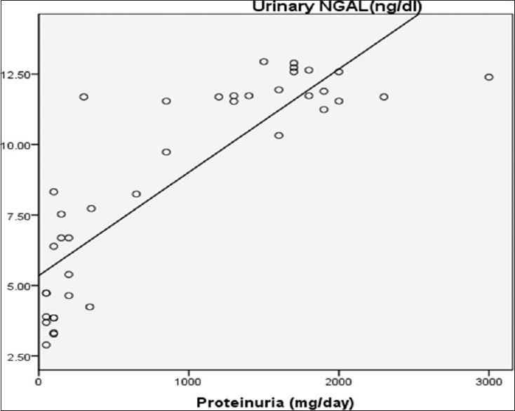
- Correlation between urinary neutrophil gelatinase-associated lipocalin and urinary protein in studied patients

- Correlation between uNGAL and Systemic Lupus Erythematosus Disease Activity Index

- Correlation between CXCL16 and urinary protein in studied patients

- Correlation between CXCL and Systemic Lupus Erythematosus Disease Activity Index in studied patients
Discussion
In the present study, we aimed to measure uNGAL and CXCL16 levels in children and adolescents with SLE and to investigate whether they are elevated in active SLE patients and to verify their role in the diagnosis of LN.
As regard to sociodemographic data of patients, female-to-male ratio was 7:1, which refers to significance of endocrinal factors in the clinical presentation of SLE. On comparison to other Arab researches in pediatric age, it was similar to the study reported by Abdel-Hafez and Abdel-Nabi, 2015[10] (7:1), lower than the study of Bakr, 2005[11] (12:1) and higher than the study of Salah et al., 2009[12] (2.7:1) and Ali et al., 1989,[13] which was on Indian kids (4:1), Uziel et al., 2007,[14] which was on Israeli children, (5:1), and Dunget et al., 2012[15] inVietnam (4:1). Difference in gender distribution might be due to different numbers of patients, due to differences in ethnicity or due to genetic factors. In general, children and adolescents have higher values of SLEDAI scores than adults.[16] Global and rSLEDAI score have been used in our study as the predictors of disease activity.[7] The mean global SLEDAI score was 28.6 ± 21.46 and was similar to that of Abdel-Hafez and Abdel-Nabi, 2015[10] (29.95 ± 2.06), and study by Dunget et al.,[15] 2012 (23.8 ± 11.6).
In the current study, there was statistically significant increase in ANA and anti-dsDNA and statistically significant decrease in C3 and C4 in the patients as compared to healthy children, this result was in agreement with Bader-Meunier et al., 2005[17] who reported almost similar results in their research.
In this study, urinary levels of NGAL and serum levels of CXCL16 were analyzed in patients with concurrent renal biopsies, which were performed at different stages during their follow-up. LN Class I was the most common finding on kidney biopsy in our study, 40 (50%) of patients, 35% was with Class III, 12.5% with Class IV, 2.5% with Class II, and no patients with Class V.
These results were not in accordance with Pluchinotta et al., 2007[18] or Ramírez et al., 2008[19] who reported that Class IV was the most dominant result in their histopathologies. The difference in the results might be attributed to different indications of kidney biopsies in different research works. Pluchinotta et al., 2007[18] and Ramírez et al., 2008[19] did biopsies only for suspected severe LN; Class I LN was of the highest proportion in our study as Class I LN patients did not have significant proteinuria or elevated serum creatinine, so we did not depend on cutoff value for proteinuria as an indication of renal biopsy and hence kidney biopsies were done in our study routinely for all newly diagnosed SLE patients even without any urinary abnormality or a rise in the level of serum creatinine.
A cohort of Suzuki et al. was the first to show an association between NGAL and diffuse proliferative glomerulonephritis in pediatric SLE patients.[20] In our study, uNGAL and sCXCL16 were significantly higher in patients with SLE than in the control groups (P < 0.05). Their levels were higher in SLE patients with renal affection than those without renal affection according to the renal biopsy results (P < 0.05). These results are consistent with theory that higher levels of uNGAL were found in patients with progressive CKD.[21] Our results were in agreement with Susianti et al. who concluded that uNGAL or combination of uNGAL and urinary transforming growth factor beta 1 or urinary monocyte chemoattractant protein-1 (uMCP-1) had the best sensitivity and specificity for active LN.[22]
In our work, uNGAL showed a higher diagnostic sensitivity (95%) and specificity (90%) than serum CXCL16 which had a sensitivity of 85% and specificity of 80% in prediction of activity of LN. Our results were in agreement with Qin et al. who reported that sCXCL16 levels were elevated in LN and recommended it to be used as serological marker of disease activity and added it might be used for evaluation of treatment strategies.[5] Singh et al. reported that CXCL16 was less promising marker for LN when compared to MCP-1 or VCAM-1.[1]
On comparing the values of uNGAL and sCXCL16 with corresponding renal histopathological classes in this study, both markers were increased in Class IV more than Class III more than Class II more than Class I, so they tended to correlate with severity of pathological findings. This was a very promising finding since Class III and IV LN have the worst outcomes, waiting intensified aggressive lines of treatment.
In this work, uNGAL and sCXCL showed statistically significant positive correlations with 24-h estimated proteinuria. The same pattern was observed when these two markers were correlated against SLEDAI disease activity scores. Since the SLEDAI indicates severe but potentially reversible inflammation, these two markers could refer to disease activity, monitoring, and thus adequate therapy. These correlations were in agreement with Brunner et al. 2006 who found a positive correlation between uNGAL and both disease activity and renal histopathological findings of kidney biopsies in their juvenile-onset SLE.[4]
Our results were in agreement with Torres-Salido et al. who evaluated fractional excretion (FE) of uNGAL relative to FE of urinary proteins ratio (FE NGAL/FE protein ratio) as it provided additional benefit to the traditional biomarkers and uNGAL to predict a proteinuric flare before worsening of rSLEDAIs and to stratify outcomes of patients with LN.[23]
In a study of Singh et al., they reported that uCXCL16 showed a prognostic value for their studied patients and added that it had a negative correlation with chronicity index as high chronicity indices indicate late-stage fibrosis and irreversible renal damage, thus unfavorable response to treatment.[1] Susianti et al. reported that NGAL and FE NGAL/FE protein ratio could predict remission following therapy, renal flaring or progression to end-stage renal disease early in the course of LN, but our results differed from their results because they reported that active LN showed significantly reduced FE NGAL/FE protein ratios, mirroring the uNGAL levels, with high specificity and sensitivity to distinguish these patients from those with nonrenal disease and from those with a previous history of treated renal involvement that remained in partial remission.[22]
Limitation of this study
One of the limitations of our work is a lack of study of validity of use of either uNGAL or sCXCL at various stages of LN (Class 1 to class 5) due to the small number of patients which was unfit for statistical analysis. What is the cutoff level to differentiate between the various classes? What is the cutoff level for the biomarkers for diagnosis of Class III/IV? The power of the study was summarized in conclusion and recommendations.
We concluded that uNGAL and CXCL16 were higher in SLE than in the control groups (P < 0.05). Their levels were higher in SLE patients with renal involvement (P < 0.05) than those without renal involvement on renal biopsys. Hence, uNGAL and CXCL16 are considered as reliable indicators of the activity of LN, which refers to the underlying histopathology of the kidney. uNGAL showed a higher diagnostic sensitivity (0.95) and specificity (0.90) than urinary CXCL16.
Financial support and sponsorship
Nil.
Conflicts of interest
There are no conflicts of interest.
References
- Urine VCAM-1 as a marker of renal pathology activity index in lupus nephritis. Arthritis Res Ther. 2012;14:R164.
- [Google Scholar]
- Prognostic factors in lupus nephritis: Diagnostic and therapeutic delay increases the risk of terminal renal failure. J Rheumatol. 2006;33:1563-9.
- [Google Scholar]
- Urinary neutrophil gelatinase-associated lipocalin as a biomarker of nephritis in childhood-onset systemic lupus erythematosus. Arthritis Rheum. 2006;54:2577-84.
- [Google Scholar]
- Elevated levels of serum sCXCL16 in systemic lupus erythematosus; potential involvement in cutaneous and renal manifestations. Clin Rheumatol. 2014;33:1595-601.
- [Google Scholar]
- Derivation and validation of the systemic lupus international collaborating clinics classification criteria for systemic lupus erythematosus. Arthritis Rheum. 2012;64:2677-86.
- [Google Scholar]
- EULAR recommendations for the management of systemic lupus erythematosus with neuropsychiatric manifestations: Report of a task force of the EULAR standing committee for clinical affairs. Ann Rheum Dis. 2010;69:2074-82.
- [Google Scholar]
- The classification of glomerulonephritis in systemic lupus erythematosus revisited. J Am Soc Nephrol. 2004;15:241-50.
- [Google Scholar]
- Statistical Methods in Medical Research Vol 4. (4th ed). Oxford: Blackwell; 2002. p. :125.
- Juvenile systemic lupus erythematosus: Onset patterns and short-term outcome in Egyptian children, a single-center experience. Lupus. 2015;24:1455-61.
- [Google Scholar]
- Epidemiology treatment and outcome of childhood systemic lupus erythematosus in Egypt. Nephrol J. 2005;20:1081-6.
- [Google Scholar]
- Outcome of a national Israeli cohort of pediatric systemic lupus erythematosus. Lupus. 2007;16:142-6.
- [Google Scholar]
- Juvenile systemic lupus erythematosus onset patterns in vietnamese children: A descriptive study of 45 children pediatric. Rheumatology. 2012;10:38-43.
- [Google Scholar]
- Difference in disease features between childhood-onset and adult-onset systemic lupus erythematosus. Arthritis Rheum. 2008;58:556-62.
- [Google Scholar]
- Initial presentation of childhood-onset systemic lupus erythematosus: A French Multicenter Study. J Pediatr. 2005;146:648-53.
- [Google Scholar]
- Distinctive clinical features of pediatric systemic lupus erythematosus in three different age classes. Lupus. 2007;16:550-5.
- [Google Scholar]
- Childhood systemic lupus erythematosus in Latin America. The GLADEL experience in 230 children. Lupus. 2008;17:596-604.
- [Google Scholar]
- Neutrophil gelatinase-associated lipocalin as a biomarker of disease activity in pediatric lupus nephritis. Pediatr Nephrol. 2008;23:403-12.
- [Google Scholar]
- Neutrophil gelatinase-associated lipocalin (NGAL) and progression of chronic kidney disease. Clin J Am Soc Nephrol. 2009;4:337-44.
- [Google Scholar]
- Analysis of urinary TGF-β1, MCP-1, NGAL, and IL-17 as biomarkers for lupus nephritis. Pathophysiology. 2015;22:65-71.
- [Google Scholar]
- Neutrophil gelatinase-associated lipocalin as a biomarker for lupus nephritis. Nephrol Dial Transplant. 2014;29:1740-9.
- [Google Scholar]


