

Translate this page into:
Hennekam lymphangiectasia syndrome
This is an open-access article distributed under the terms of the Creative Commons Attribution-Noncommercial-Share Alike 3.0 Unported, which permits unrestricted use, distribution, and reproduction in any medium, provided the original work is properly cited.
This article was originally published by Medknow Publications and was migrated to Scientific Scholar after the change of Publisher.
Abstract
Hennekam lymphangiectasia syndrome is a rare disorder comprising of intestinal and renal lymphangiectasia, dysmorphic facial appearance and mental retardation. The facial features include hypertelorism with a wide, flat nasal bridge, epicanthic folds, small mouth and small ears. We describe a case of a multigravida with bad obstetric history and characteristic facial and dental anomalies and bilateral renal lymphangiectasia. To our knowledge this is the first case of Hennekam lymphangiectasia syndrome with anodontia to be reported from India.
Keywords
Facial and dental anomalies
renal lymphangiectasia
Hennekam lymphangiectasia syndrome

Introduction
Hennekam Lymphangiectasia syndrome is a rare disorder characterized by presence of intestinal and renal lymphangiectasia, dysmorphic facial appearance and mental retardation. The facial features include hypertelorism with a wide, flat nasal bridge, epicanthic folds, small mouth and small ears. This syndrome thought to be due to developmental disorder of the lymphatics. The synonym for this disease is multiple congenital anomaly/mental retardation (MCA/MR) syndrome.
Case Report
A 38-year-old female with previous bad obstetric history came to our institution with 30 weeks of amenorrhea. She married 12 years back (non-consanguineous) and had four pregnancies prior to the present one. The first pregnancy was 6 months after marriage; it was an ectopic pregnancy and was terminated. The second pregnancy was 2 months later, terminated at 8 weeks as she was diagnosed to have a “renal disease”; the third was 2 years later, had an intrauterine death at six and half months of gestation; in the 4th pregnancy the fetus was found to have multiple congenital anomalies (MCA) (hydrocephalus, flat nasal bridge and club foot), was delivered vaginally at 6 months of gestation and it was stillbirth. She was diagnosed as having “renal parenchymal disease” during the second pregnancy and was told to have bilateral enlarged kidneys. She also gives history of non-eruption of all permanent teeth and has had surgery for umbilical hernia repair in 2006. The hernia was probably due to raised intra-abdominal pressure secondary to enlarged kidneys.
She had noticed frothing of urine and increasing pedal edema at 20 weeks of gestation in the present pregnancy. There was no history oliguria, macrohematuria or lithuria. There was no history of skin rash, photosensitivity, oral ulcers, behavioral abnormalities or pain/discoloration of limbs. There was no history of NSAIDs or native medicine intake. There was history of renal disease or similar illness in the family.
On examination, she had anasarca, pallor and blood pressure was 148/94 mmHg. General examination showed facial and dental anomalies that included flat face, flat nasal bridge, hypertelorism [Figure 1a], epicanthal folds, small mouth, low set ears [Figure 1b] and absent teeth (anodontia) [Figure 1c]. There was no growth retardation or MR. Systemic examination and gynecological evaluation were normal and uterus was enlarged corresponding to 28 weeks and fetal heart sounds were present.
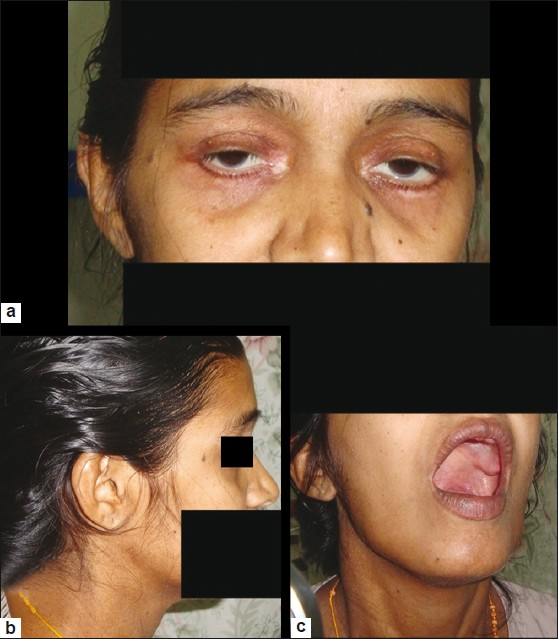
- Photograph showing typical facial and dental anomalies of Hennekam lymphangiectasia syndrome – (a) depressed nasal bridge, hypertelorism (b) low-set ears and (c) anodontia
Investigations revealed 2+ proteinuria and microhematuria, severe hypoalbuminemia (1.5 g/dl) and hypoglobulinemia (2.5 g/dl). Her serum creatinine was 1.0 mg/dl; hemoglobin was 10.2 g/dl; had lymphocytopenia (890 cells/cu.mm), platelet and other leukocyte counts were normal. Ultrasonogram of abdomen showed bilateral perinephric fluid collection; kidneys were normal sized with preserved cortico-medullary differentiation. The fetus had evidence of IUGR with gestational age of 27 weeks on ultrasound evaluation and there were no congenital anomalies. She had an MDCT of abdomen taken prior to conception, which showed fluid collections (fluid attenuation was 0-10 HU, this differentiates lymph from any soft tissue masses) in the perinephric and peripelvic space surrounding the renal cortex bilaterally (Figure 2a – transverse and Figure 2b – coronal sections) which is characteristic of renal lymphangiectasia.

- MDCT images showing fluid collections in the perinephric and peripelvic space bilaterally, surrounding the renal cortex (a – transverse and b – coronal sections) suggestive of renal lymphangiectasia – (black arrows – perinephric collection and white arrows – kidneys)
The blood pressure was controlled with two antihypertensive drugs (Nifedipine 10 mg twice daily and α-Methyl Dopa 500 mg thrice daily). Her edema was controlled with fresh frozen plasma transfusions and parentral diuretics. She underwent emergency LSCS as she developed oliguria and mild renal insufficiency (1.9 mg/dl); a preterm male child of 600 g was delivered and was shifted to neonatal ICU for resuscitation. The baby expired 14 days after delivery due to prematurity and neonatal sepsis. The baby did not have any congenital anomalies and the postnatal USG did not show any perinephric collection in the newborn. Antihypertensive medication could be reduced after the delivery and she had improvement in urine volume and normalization of serum creatinine (1.1 mg/dl). She was discharged with an advice to come for follow-up for monitoring hypertension, proteinuria and renal parameters.
She was diagnosed to Hennekam lymphangectasia syndrome in view of characteristic facial and dental anomalies (flat face, flat nasal bridge, hypertelorism, epicanthal folds, small mouth, absent tooth and low set ears), hypoalbuminemia and hypoglobulinemia and lymphocytopenia (suggestive of intestinal lymphangiectasia). The ultrasonographic and MDCT findings in her are characteristic of this disease.
Discussion
Hennekam Raoul, a Dutch physician, described a syndrome of intestinal lymphangiectasia with severe lymphedema of the limbs, genitalia and face and severe MR in 1989.[1] This syndrome was thought to be due to developmental disorder of the lymphatics. The synonym for this disease is MCA/ MR syndrome. The additional features of the syndrome described in various case reports are facial and dental anomalies like flat face, flat nasal bridge, hypertelorism, epicanthal folds, flat upper lip, small mouth, narrow palate, mild retrognathia, craniosynostosis, dysmorphic pinnae, atresia of ear canal, oligodontia, and conical crowns. The dermatological and other appendagial anomalies described are severe lymphedema of the limbs, genitalia and face; infection of oozing lymphatics (erysipelas), alopecia areata, frontal upsweep and heavy eyebrows. Other systemic features reported are of thorax (narrow upper thorax), cardiovascular system (ventricular septal defect) and central nervous system (severe mental retardation, seizures).
The prominent anomaly reported with gastrointestinal tract is intestinal lymphangiectasia, accompanied usually with hypoproteinemia, hypogammaglobulinemia, and lymphocytopenia. Urogenital anomalies described are genital lymphedema, kidney malformations, and vesicoureteral reflux.
Following Hennekam's initial descriptions of the syndrome, there are multiple reports of the same all over the World;[2–9] to our knowledge, this is first case to be reported from India.
The syndrome is familial and was first reported in two male and two female children of consanguineous parents.[1] The pattern of transmission was autosomal recessive.[1] It seems likely that most (but not all) manifestations of the entity can be explained as sequences of impaired prenatal and postnatal lymphatic flow, suggesting that the causative gene(s) should have a major function in lymphangiogenesis. Among the important candidate genes for analysis, Prox-1 is being investigated.[9] Prox-1 is expressed initially in a subpopulation of the vascular endothelial cells at the sites where the lymphatics bud off the veins and later becomes confined to the lymphatic endothelial cells.[9] Prox-1 is required for both emergence of lymphatic endothelial cells from the veins and their differentiation towards the lymphatic phenotype.[9] Histology shows malformation or dilation of lymphatic channels resulting in lymph blockages and accumulation of fluids occur in the affected body areas.
Alders et al. have reported seven patients with autosomal recessive Hennekam syndrome. They have identified a critical chromosomal region containing CCBE1 (collagen and calcium binding EGF domains 1), the human ortholog of a gene essential for lymphangiogenesis in zebrafish by homozygosity mapping. Homozygous and compound heterozygous mutations in these subjects paired with functional analysis in a zebrafish model identify CCBE1 located on long arm of Chromosome 18 (18q21.32) as one of few genes causing primary generalized lymph-vessel dysplasia in humans.[10]
The condition usually presents as facial and dental anomalies. Facial features are thought to mirror the extent of intrauterine facial lymphedema, or may be caused by lymphatic obstruction that affects the early migration of neural crest tissue. Other associated findings are varying degree of MR and occasionally seizures. The patients may have lymphedema with or without cellulitis. Intestinal or pleural lymphangiectasia results in hypoproteinemia and edema. Renal lymphangiectasis is most often asymptomatic and detected on routine imaging and is one of the differential diagnoses for bilateral enlarged palpable kidneys. Renal lymphangiectasis is characterized by presence of fluid collections (with very low attenuation of 0-10 HU, this differentiates lymph from any soft tissue masses) in the perinephric, peripelvic space bilaterally, surrounding the renal cortex on MDCT. There is no curative treatment for lymphangiectasia. Treatment is focused on control of complications like hypoproteinemia, cellulitis and seizures.
Hennekam lymphangiectasia syndrome is a rare disorder comprising of dysmorphic facial appearance, MR, intestinal and renal lymphangiectasia. Treatment is not usually necessary, as it does not affect the renal function. Our patient is the first case to be reported from India and has anodontia, which has not been described before.
Source of Support: Nil
Conflict of Interest: None declared.
References
- Autosomal recessive intestinal lymphangiectasia and lymphedema, with facial anomalies and mental retardation. Am J Med Genet. 1989;34:593-600.
- [Google Scholar]
- Craniosynostosis and kidney malformation in a case of Hennekam syndrome. Am J Med Genet. 1995;57:66-8.
- [Google Scholar]
- Intestinal lymphangiectasia, lymphedema, mental retardation, and typical face: confirmation of the Hennekam syndrome. Am J Med Genet. 2005;40:244-7.
- [Google Scholar]
- Cutaneous manifestations and massive genital involvement in Hennekam syndrome. Pediatr Dermatol. 2006;23:239-42.
- [Google Scholar]
- Lymphedema-lymphangiectasia-mental retardation (Hennekam) syndrome: A review. Am J Med Genet. 2002;112:412-21.
- [Google Scholar]
- Hennekam Lymphangiectasia-Lymphedema Syndrome. Centre for Arab Genomic Studies, A Division of Sheikh Hamdan Award for Medical Sciences. The Catalogue for Transmission Genetics in Arabs, CTGA Database. :1-2.
- [Google Scholar]
- Mutations in CCBE1 cause generalized lymph vessel dysplasia in humans. Nat Genet. 2009;41:1272-4.
- [Google Scholar]


