

Translate this page into:
Takayasu arteritis in an infant
This is an open-access article distributed under the terms of the Creative Commons Attribution-Noncommercial-Share Alike 3.0 Unported, which permits unrestricted use, distribution, and reproduction in any medium, provided the original work is properly cited.
This article was originally published by Medknow Publications & Media Pvt Ltd and was migrated to Scientific Scholar after the change of Publisher.
Abstract
Takayasu arteritis (TA), a chronic inflammatory arteritis affecting the aorta and its main branches, is a rare condition mainly affecting young women in the second and third decades of life. Occurrence of TA in infants is extremely rare, with only less than 10 cases reported all over the world until date. We report a case of a 2-year-old girl who presented with hypertension and was diagnosed to have TA with bilateral renal artery stenosis and this is probably the youngest case reported from India.
Keywords
Childhood vasculitis
hypertension in infants
renovascular hypertension
takayasu arteritis

Introduction
Takayasu arteritis (TA) is a chronic inflammatory arteritis affecting the aorta and its main branches. The disease has a world-wide distribution, but has a high prevalence in Japan.[1] Although there are no population based studies from India, reports have shown than the prevalence may be as high as in Japan.[23] Most of the patients diagnosed to have this condition are women in their second and third decades of life.[4] The occurrence of the disease in young children and infants is extremely rare with only a few cases reported all over the world.[567] We report a case of TA in an infant.
Case Report
The present case report is about a 2-year-old female patient who was brought to the hospital with a history of incidentally detected hypertension of 2 months duration. She was born out of a non-consanguineous marriage and had an uneventful antenatal and postnatal course with normal developmental milestones and she had received vaccinations according the National Immunization Program, which includes Bacillus calmette-guerin (BCG). On examination, she was afebrile, active and well-nourished; her weight was 16 kg and height was 80 cm. Blood pressure was 140/90 mmHg, which was more than the 99th percentile for her height. Blood pressure was equal in all four limbs. All peripheral pulses were palpable. Fundus examination showed mild arteriolar narrowing on both sides. Systemic examination was unremarkable. There was no abdominal bruit. Her renal functions were normal (blood urea: 8.4 mg/dl; serum creatinine: 0.39 mg/dl). Serum sodium was 133.5 mmol/l, S. potassium 3.3 mmol/l, S. calcium 9.9 mg/dl and S. phosphurus 3.94 mg/dl. Urine examination showed 1 + proteinuria and microscopy was normal. Erythrocyte sedimentation rate (ESR) was 20 mm/1 h and C-reactive protein was 6 mg/dl. X-ray chest and electrocardiography were normal. Ultrasonogram of abdomen revealed asymmetric kidneys, with a smaller left kidney. The size of the right kidney was 7.1 cm × 3.6 cm and that of the left kidney was 6.1 cm × 2.9 cm. Computed tomography aortogram showed critical stenosis of the left renal artery, at a length of 8 mm from the origin, a short segment stenosis of the right renal artery at its origin, a tight focal stenosis at the origin of the celiac artery with post-stenotic dilatation, mild stenosis at the origin of the superior mesenteric artery and narrowing of the abdominal aorta at the level of renal arteries; there was no para-aortic lymphadenopathy [Figure 1]. The diameter of aorta at the level of diaphragm, renal arteries and just above bifurcation into iliac arteries were 6.2mm, 5.2 mm and 6.6 mm respectively, proving that there was a narrowing at the level of renal arteries followed by post-stenotic dilatation. With these angiographic findings, in this infant with hypertension, a diagnosis of TA was made according to the European League Against Rheumatism/Pediatric Rheumatology International Trials Organization/Pediatric Rheumatology European Society (EULAR/PRINTO/PRES) criteria - 2008.[8] Angioplasty of the left renal artery was attempted, but was not successful. The child is being treated with nifedipine 5 μg thrice daily and clonidine 50 mg thrice daily and her blood pressure is 90/55 mmHg.

- Computed tomography angiogram showing bilateral renal artery stenosis and narrowing of abdominal aorta (arrows: A: Short segment stenosis of right renal artery, B: Stenosis of left renal artery with small left kidney, C: Narrowing of the abdominal aorta at the level of renal arteries)
Discussion
TA, also known as “pulseless disease”, “occlusive thromboaortopathy” and “Martorell syndrome”, was first systematically described by a Japanese Ophthalmologist Mikito Takayasu. The disease remains an enigma as the exact cause of the disease is still not elucidated. Hypersensitivity to Mycobacterium tuberculosis has been postulated to be an important etiological factor.[9] A Human Leucocyte Antigen (HLA) association and viral etiology have also been postulated.[10]
The clinical presentation of TA includes three phases:
-
The early phase or prepulseless phase characterized by nonspecific systemic features such as malaise, arthralgia, weakness, weight loss and low grade fever
-
The pulseless phase characterized by claudication, amaurosis or diplopia and renovascular hypertension
-
The occlusive phase characterized by transient ischemic attack, stroke, aortic regurgitation, cardiac failure, renovascular hypertension and claudication.
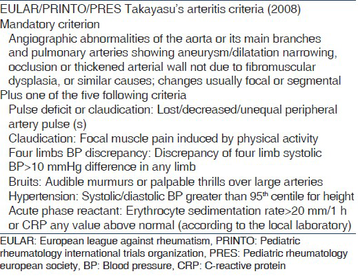
The diagnosis of TA in children is based on the EULAR/PRINTO/PRES criteria[8] [Table 1].
TA is certainly underdiagnosed and underreported from India, due to the fact that the disease may not present with classical features and the imaging modalities are available only in tertiary care centers. A study that compared the clinical manifestations of TA in India and Japan reported that there was a significant variation in the manifestations of the disease in the two countries.[4] In contrast to Japanese patients in whom proximal aorta involvement (Takayasu conference classification - type I and II) was common, in Indian patients descending and abdominal aorta involvement (type IV) was common. Moreover, hypertension was the most dominant sign in Indians compared with pulse lessness in the Japanese. It was also noted that the age at diagnosis of the disease was almost a decade earlier in Indians compared to Japanese (28 ± 10 vs. 37 ± 14 years, P < 0.01). In an Indian study, it was found that aortoarteritis was the most common cause of renovasular hypertension and accounted for 59.4% of all cases. However, the mean age of the subjects was 27 years and the youngest child was 5 years of age.[11]
To the best of our knowledge, there are no reports from India of a case of TA in an infant. Our patient had type IV disease, with the involvement of abdominal aorta, coeliac, superior mesenteric and both renal arteries. Although it is suggested that glucocorticoids are the mainstay of treatment in the early or prepulseless phase, other immunosuppressive agents like Methotrexate, Azathioprine and anti-tumor necrosis factor agents have also been used, especially in steroid resistant cases. However, no evidence is available for benefit of therapy in the pediatric age group. Angioplasty is preferred for stenotic lesions amenable to catheter-based therapy. However percutaneous intervention is less likely to succeed when stenosis or occlusions affect lengthy portions of an artery or the artery is heavily scarred. An angioplasty to the left renal artery was attempted in this case, but was unsuccessful. Hence it was decided to continue medical management with anti-hypertensives as blood pressure could be controlled with medications and she was not given steroids as there were no clinical or laboratory indicators of an active inflammatory disease in this infant.
Conclusion
Although the world-wide prevalence of TA is very low, it is more prevalent in Asian countries like Japan and India. The clinical manifestations of the disease in India are different from that seen in Japan. Type IV disease involving abdominal aorta and renal arteries is more common in Indians and hypertension is the most common sign. If the disease is active, steroids can be tried and the lesions amenable to angioplasty may be corrected by a percutaneous transluminal angioplasty. TA is extremely rare in infancy. To the best of our knowledge, there are no case reports of TA in an infant from India. This is probably the youngest patient reported to have TA in India.
Source of Support: Nil
Conflict of Interest: None declared.
References
- Current status of aortoarteritis in India. J Assoc Physicians India. 2004;52:48-52.
- [Google Scholar]
- Clinical manifestations of Takayasu arteritis in India and Japan – New classification of angiographic findings. Angiology. 1997;48:369-79.
- [Google Scholar]
- Case report of a 2-year-old boy with Takayasu's arteritis: An atypical, severe presentation of a rare disease. Pediatr Cardiol. 2010;31:1089-92.
- [Google Scholar]
- Sudden death of an infant with coronary involvement due to Takayasu arteritis. Cardiovasc Pathol. 2013;22:109-11.
- [Google Scholar]
- Aortic valve replacement surgery for a case of infantile Takayasu arteritis. Korean J Pediatr. 2012;55:254-8.
- [Google Scholar]
- EULAR/PRINTO/PRES criteria for Henoch-Schönlein purpura, childhood polyarteritis nodosa, childhood Wegener granulomatosis and childhood Takayasu arteritis: Ankara 2008. Part II: Final classification criteria. Ann Rheum Dis. 2010;69:798-806.
- [Google Scholar]
- Spectrum of renovascular hypertension in the young in north India: A hospital based study on occurrence and clinical features. Angiology. 1985;36:370-8.
- [Google Scholar]


