

Translate this page into:
Fusarium solani infection in a kidney transplant recipient
This is an open-access article distributed under the terms of the Creative Commons Attribution-Noncommercial-Share Alike 3.0 Unported, which permits unrestricted use, distribution, and reproduction in any medium, provided the original work is properly cited.
This article was originally published by Medknow Publications & Media Pvt Ltd and was migrated to Scientific Scholar after the change of Publisher.
Abstract
Hyalo hypho mycosis due to Fusarium species mainly occurs in immunocompromised hosts. The clinical presentation varies from localized to disseminated involvement. A case of localized cutaneous fusariosis caused by Fusarium solani in a renal transplant patient is described and the skin manifestations of the disease are discussed.
Keywords
Fusarium
Fusariosis
transplantation

Introduction
Fusariosis is a rare infectious disease caused by species of the genus Fusarium that has been increasingly documented as an emerging agent of opportunistic infections in immunocompromised patients. Fusarium species are molds that are distributed worldwide and that may be recovered from a wide range of substrates. Portals of entry include the respiratory and the gastrointestinal tracts, catheter tips, indwelling central venous catheters and the skin. Patients with cutaneous diseases related to Fusarium species can present with superficial and deep infections as well as toxic reactions. In addition, Fusarium may colonize in wounds. There are several ways the cutaneous Fusarium infection manifests, but commonly seen with erythematous papules and nodules with central necrosis and subcutaneous nodular lesions.
Case Report
A 42-year-old man, who had live related donor renal transplantation 18 years ago, and was taking azathioprine 100 mg and prednisolone 10 mg once daily as immunosuppesive agents, developed new onset diabetes mellitus 6 months after transplantation and was on insulin since then. He has a stable graft function without any rejection episode. Three and half years ago he developed a small ulcer on the skin over right knee joint which was operated, after which he had remained asymptomatic for 3 years. He then developed papulo-nodular lesions and erythematous plaques with multiple discharging sinuses at that site which had developed after a minor trauma. These lesions gradually evolved to multiple discharging sinuses [Figure 1]. Prior to his visit to us he consulted surgeon to remove the ulcers. The surgeon operated thrice but after each surgery the sinuses and ulcer increased in number and size. On examination, there were scattered erythematous papules and nodules with necrotic ulcerations and multiple discharging sinuses. There was no lymphadenopathy. Histopathology revealed a granulomatous suppurative infiltrate extending to the entire dermis and numerous ectatic blood vessels. Many hyaline hyphae and unicellular fungal elements were seen in the infiltrate and within the vascular spaces. Gomori Methenamine staining showed hyphae of more than 1μm in diameter and reproductive structures represented by microconidia and chlamydospore like structures, suggesting a presumptive diagnosis of non-Aspergillus hyalo-hypho-mycosis. Sabouraud-Dextrose agar culture without cycloheximide grew whitish grey cottony colonies suggestive of Fusarium species. Successive subcultures performed on potato dextrose agar, then put on lactophenol cotton blue mount which showed sickle-shaped multiseptatedmacroconidia; and one-to two-celled microconidia formed from unbranched phialides, conidiophores and chlamydospores typical of Fusarium [Figures 2 and 3]. Laboratory investigations revealed impaired renal function, with raised serum creatinine levels (2.8 mg/dl) and blood urea levels (87 mg/dl). Three consecutive blood cultures were negative. X-ray of the chest and gastrointestinal tract, as well as an abdominal ultrasound, showed no abnormalities. Itraconazole was started at 100 mg twice daily, which led to after 6 weeks.
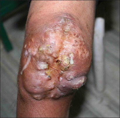
- Multiple discharging sinuses

- Acutely branched fungal hyphae seen on GMS stain (×40)

- Acutely branched fungal hyphae seen on GMS stain (×100)
Discussion
Fusarium species are important plant pathogens causing a broad spectrum of infections in humans, including superficial (such as keratitis and onychomycosis), locally invasive, or disseminated infections which occur almost exclusively in immunocompromised patients particularly those with prolonged and profound neutropenia and/or severe T-cell immunodeficiency.[123] Locally invasive and late infections may also develop among solid organ transplant recipients,[4] but these appear to be less common than those among HSCT patients.
The principal portal of entry for Fusarium is the airway, followed by the skin at the site of tissue breakdown and the mucosal membranes. Airborne fusariosis is acquired by the inhalation of airborne fusarial conidia as suggested by the occurrence of sinusitis and or pneumonia in the absence of dissemination. Skin as a portal of entry is supported by the development of infection following skin breakdowns due to trauma (automobile accidents, bomb injury), burns or onychomycosis in normal hosts[5] and the development of cellulitis (typically at sites of tissue breakdown such as toes and fingers), which may remain localized or lead to disseminated infection in immunocompromised patients.[26]
Skin involvement can represent a primary site of infection, usually a cellulitis of the toes, or a manifestation of metastatic infection in patients with disseminated fusariosis. Skin involvement in fusariosis was present in 181 patients (70%) among 259 published cases of fusariosis (232 immunocompromised and 27 immunocompetent).[5] Among immunocompetent hosts, lesions are usually localized (13 of 14 patients) and occur after skin breakdown (trauma or pre-existing onychomycosis).[5] Among immunocompromised patients, skin lesions may also be localized, usually as a result of skin breakdown caused by trauma, or may lead to disseminated infection. Among 16 patients with metastatic skin lesions, a recent history of cellulitis at the site of onychomycosis (11 patients), local trauma (3 patients), or an insect bite (2 patients) was reported.[5] Patients with disseminated disease typically have multiple erythematous papular or nodular and painful lesions, frequently with central necrosis giving the lesions an ecthyma gangrenosum like appearance. Target lesions (a thin rim of erythema of 1-3 cm in diameter surrounding the above-mentioned papular or nodular lesions) may be present in approximately 10% of patients while bullae develop rarely. Fusarial skin lesions can involve practically any site, with predominance in the extremities and evolve rapidly, usually over a few days. Lesions at different stages of evolution (papules, nodules and necrotic lesions) may be present in a third of patients and concomitant myalgia (suggesting muscle involvement) was described in 15%. Skin lesions were the single source of diagnosis in the majority of patients with such lesions 100/181[55%].[5]
The diagnosis of Fusarium infection is principally based on mycology and histopathology. Recently, polymerase chain reaction technique has also been developed for specific detection of Fusarium species from both culture and clinical samples.[7] Cultures require incubation at 25°C on a Sabouraud Dextrose medium without cycloheximide. The most important microscopic features in species identification on culture are the conidia: The presence of fusoidmacroconidiain which there are foot cells with some type of heel is accepted as the most definitive characteristic of the genus Fusarium.[8] Histologically, diagnostic clues include the presence of adventitious sporulation consisting of phialides and phialoconidia and the presence of irregular hyphae with both 45° and 90° branching in a closed lesion.[910] Although culture remains the standard for identification of these fungi, presumptive histological identification of non-Aspergillus hyalohyphomycoses, as in our case, is of great value for several reasons. In fact, as suggested by Liuetal.,[9] histopathologic evidence of hyalohyphomycoses is helpful when culture is not requested or is unsuccessful for technical reasons. Moreover, histology results can be obtained in <24 h, leading to the prompt institution of therapy. This is of importance because of both the rapid dissemination of infection in immunocompromised hosts and the frequent resistance of Fusarium species to antifungal drugs.
Conclusion
Fusarium is a rare fungal infection in immunocompormised host needs good clinical suspicion and prompt identification and confirmation by mycology and histopathology for its management. Surgical management should be considered only in select cases when all sort of conservative management fails. Most of the antifungals available at present are not effective.
Source of Support: Nil
Conflict of Interest: None declared.
References
- Fusarium, a significant emerging pathogen in patients with hematologic malignancy: Ten years’ experience at a cancer center and implications for management. Blood. 1997;90:999-1008.
- [Google Scholar]
- Fusarium infection in hematopoietic stem cell transplant recipients. Clin Infect Dis. 2004;38:1237-42.
- [Google Scholar]
- Fusarium infection after solid-organ transplantation. Clin Infect Dis. 2001;32:1237-40.
- [Google Scholar]
- Cutaneous infection by Fusarium species in healthy and immunocompromised hosts: Implications for diagnosis and management. Clin Infect Dis. 2002;35:909-20.
- [Google Scholar]
- Outcome predictors of 84 patients with hematologic malignancies and Fusarium infection. Cancer. 2003;98:315-9.
- [Google Scholar]
- Specific detection of Fusarium species in blood and tissues by a PCR technique. J ClinMicrobiol. 1999;37:2434-8.
- [Google Scholar]
- Taxonomy, biology, and clinical aspects of Fusarium species. ClinMicrobiol Rev. 1994;7:479-504.
- [Google Scholar]
- Morphologic criteria for the preliminary identification of Fusarium, Paecilomyces, and Acremonium species by histopathology. Am J ClinPathol. 1998;109:45-54.
- [Google Scholar]
- Morphologic identification of mycelial pathogens in tissue sections. A caveat. Am J ClinPathol. 1998;109:1-2.
- [Google Scholar]


