

Translate this page into:
Unmasking of complements using proteinase-K in formalin fixed paraffin embedded renal biopsies
This is an open access article distributed under the terms of the Creative Commons Attribution-NonCommercial-ShareAlike 3.0 License, which allows others to remix, tweak, and build upon the work non-commercially, as long as the author is credited and the new creations are licensed under the identical terms.
This article was originally published by Medknow Publications & Media Pvt Ltd and was migrated to Scientific Scholar after the change of Publisher.
Abstract
Renal biopsy interpretation requires histopathology, direct immunofluorescence (DIF) and electron microscopy. Formalin-fixed, paraffin-embedded tissue (FFPE) sent for light microscopy can be used for DIF after antigen retrieval. However, complement staining has not been satisfactory. We standardized DIF using proteinase-K for antigen retrieval in FFPE renal biopsies. A pilot study was conducted on known cases of membranous glomerulonephritis (MGN), membranoproliferative type-1 (MPGN-1), immunoglobulin A nephropathy (IgAN), and anti-glomerular basement disease (anti-GBM). Immunofluorescence panel included fluorescein isothiocyanate (FITC) conjugated IgG, IgA, IgM, complements (C3 and C1q), light chains (kappa, lambda) and fibrinogen antibodies. After standardization of the technique, 75 renal biopsies and 43 autopsies cases were stained. Out of 43 autopsy cases, immune-complex mediated glomerulonephritis (GN) was confirmed in 18 cases (Lupus nephritis-11, IgAN-6, MGN-1), complement-mediated dense deposit disease (DDD-1) and monoclonal diseases in 4 cases (amyloidosis-3, cast nephropathy-1). Immune-mediated injury was excluded in 17 cases (focal segmental glomerulosclerosis -3, crescentic GN-6 [pauci-immune-3, anti-GBM-3], thrombotic microangiopathy-5, atherosclerosis-3). Renal biopsies (n-75) where inadequate or no frozen sample was available; this technique classified 52 mesangiocapillary pattern as MPGN type-1-46, DDD-2 and (C3GN-4). Others were diagnosed as IgAN-3, lupus nephritis-2, MGN-4, diffuse proliferative glomerulonephritis (DPGN)-1, Non-IC crescentic GN-1, monoclonal diseases-3. In nine cases, DIF on FFPE tissue could not help in making diagnosis. Proteinase-K enzymatic digestion of FFPE renal biopsies can unmask complements (both C3 and C1q) in immune-complexes mediated and complement-mediated diseases. This method showed good results on autopsy tissues archived for as long as 15 years.
Keywords
Antigen retrieval
complement
direct immunofluorescence
formalin-fixed paraffin-embedded
membranoproliferative
proteinase-K

Introduction
Renal biopsy interpretation requires histological examination by light microscopy, direct immunofluorescence (DIF)/immunohistochemistry, and electron microscopy. DIF is simple, fast and very sensitive technique in which fluorescent tagged monoclonal antibodies are used for staining on frozen kidney biopsies sections. Sometimes kidney biopsy core processed for DIF do not contain any glomeruli. In such cases, Formalin-fixed, paraffin-embedded (FFPE) tissue sections prepared from the portion of a biopsy sent for light microscopy can be used as substrate for DIF. In such case, immunofluorescence (IF) can be performed by using different antigen retrieval methods with proteases such as pepsin, trypsin, or other enzymes.[12] These work well for most of the immunoglobulins (Igs) and light chains but do not give good results for complements.
We substituted these enzymes by proteinase-K for antigen retrieval and followed the rest of the procedure as originally followed by Fogazzi et al.,[3] and Nasr et al.,[4] Proteinase-K is frequently used in molecular laboratories for DNA isolation, protein digestion experiments and is easily available in most laboratories. After satisfactory standardization on controls cases with a known diagnosis, we performed DIF on 75 cases of paraffin-embedded renal biopsies in which no glomeruli were present for interpretation on fresh IF core or no separate tissue was available. Similarly, kidney sections of 43 cases of autopsy with the morphologic suggestion of renal disease were analyzed using proteinase-K antigen retrieval. DIF could be accomplished using proteinase-K antigen retrieval method and has the advantage of complement unmasking that was not possible with previously described methods.
Materials and Methods
This study was carried out on the control set comprising of known cases of membranous glomerulonephritis (MGN), (n-11), membranoproliferative (MPGN)-type-1 (n-11), IgA nephropathy (IgAN) (n-9) and anti-glomerular basement disease (anti-GBM) disease (n-2). After getting comparable results, a study set comprising of 75 renal biopsies (stored up to 5 years) and 43 autopsies (stored up to 15 years) were used. IF staining performed after antigen retrieval using proteinase-K on FFPE tissues. On poly-L-lysine coated slides, 3µm sections were taken. Slides were kept at 37°C overnight. Deparaffinization was done by giving two changes in xylene for 10 min each, one change in ethanol 100% for 5 min followed by changes in 70% and 50% alcohol for 5 min. Slides were rehydrated by washing in distilled water (20 dips) and kept in Tris buffer (pH 9.0) at 37°C for 20 min. Incubation with proteinase-K, (Amresco, OH 44139 USA, Cat 0706),) 0.25 mg/ml was done at 37°C for 5 min. After washing with Tris buffer, (pH-9.0) slides were incubated at 4°C for 20 min. Incubation was performed for 2 h in a wet humidified chamber. fluorescein isothiocyanate (FITC) conjugated polyclonal antibodies directed for immunoglobulins (IgG, IgA, IgM), complements (C3, C1q), light chains (kappa, lambda) and fibrinogen (Dako, Carpinteria, CA, USA) were used as per manufacturer instructions. Finally, slides were rinsed in PBS, and aqueous mounting with phosphate buffer glycerine was done. Slides were examined under a dark field ultraviolet microscope. DIF intensity was scored semi-quantitatively on a scale of 0 (negative), 0.5+ (trace), 1+ (mild), 2+ (moderate) and 3+ (strong positivity) and compared with DIF interpretation on fresh tissue in control set. This study was approved by the institute's ethics committee.
Results
After comparing the staining intensities on fresh and paraffin-embedded biopsies we observed the same staining patterns of Igs and complements, however, intensity of the score was one grade less in most cases. The granular texture was less appreciable, and the staining pattern was segmental and focal in some cases. Staining in anti-GBM disease was negative to trace [Figure 1].
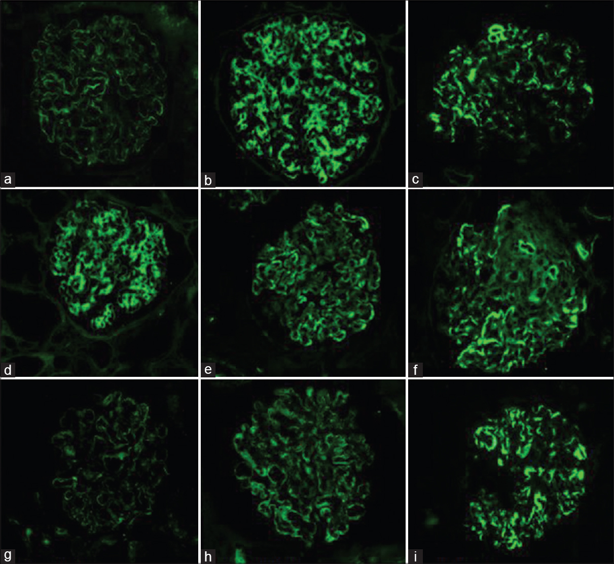
- Photomicrographs shows, (a) patchy glomerular basement membranes staining (2+), (b) mesangial (3+) staining with immunoglobulins (IgG), (c) granular staining for IgG, (d) IgA in mesangium (3+), (e) IgM (3+) staining of glomerular capillary loops and mesangium, (f) Granular staining for C3 (3+), (g) linear kappa staining (2+) in glomerular capillary loops and mesangium, (h) lambda staining (3+) for glomerular capillary loops, (i) granular staining for C1q in membrane and mesangium. Note: No background staining observed, but granularity is less appreciable (fluorescein isothiocyanate [a-i, ×20])
After the standardization of this technique, 75 renal biopsies were analyzed by DIF using proteinase-K. Fifty two cases had mesangiocapillary pattern and categorized as MPGN Type 1 (n-46), dense deposit disease (DDD) (n-2) and C3GN (n-4). Others were diagnosed as IgAN (n-3), lupus nephritis (n-2), MGN (n-4), diffuse proliferative glomerulonephritis (DPGN) (n-1), Non-IC crescentic GN (n-1), monoclonal diseases (n-3), light chain cast nephropathy (n-1), monoclonal Ig deposition disease (n-1) and proliferative GN with monoclonal Ig deposition (n-1). In nine cases, DIF on FFPE tissue could not help in making a diagnosis [Table 1].

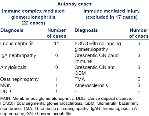
Out of 43 autopsy cases, immune-complex mediated GN was confirmed in 18 cases (Lupus nephritis-11, IgAN-6, MGN-1). Complement-mediated diseases like DDD (n-1) and monoclonal disease was diagnosed in 4 cases (amyloidosis-3, cast nephropathy-1) [Table 2]. Immune-mediated injury excluded in 17 cases (focal segmental glomerulosclerosis [FSGS] with collapsing glomerulopathy-3, crescentic GN-6 (pauci immune-3, anti-GBM disease-3), Thrombotic microangiopathy-5, atherosclerosis-3. We had good staining with complements, that is, C3 and C1q in all possible patterns with different diagnosis in paraffin sections [Figure 2].

- Photomicrographs shows C3 staining, (a) C3 mesangial staining, (b) granular (arrow) staining along glomerular capillaries in membranous glomerulonephritis (GN), (c) coarse mesangial (2+) focal extending to capillaries in dense deposit disease, (d) peripheral glomerular capillaries as seen in C3 GN, (e) C1q staining glomerular capillaries which is segmentally negative, (f) C1q along tubular basement membrane (fluorescein isothiocyanate [a, c-f: ×20, b: ×40])
Two autopsy cases with the diagnosis of MGN and lupus nephritis class IV, for which previous IF was available, are shown to compare the intensity of staining [Figures 3 and 4]. There were certain differences in the staining intensities when we performed IF on FFPE sections. We found that the intensity on FFPE was one score less than that seen on fresh tissues [Table 3]. Staining was also patchy in certain cases. However, background was absent, and pattern of staining was less granular than seen on fresh tissue with smudgy appearance in some cases.

- Photomicrographs showing comparison of immunofluorescence with full panel on fresh tissue and paraffin section of the same case with diagnosis of membranous glomerulopathy (fluorescein isothiocyanate tagged antibodies used immunoglobulins, C3, Kappa, Lambda, ×20)

- Photomicrohraphs showing comparison of immunofluorescence done on the full panel of fresh tissue and paraffin section of the same case with diagnosis of lupus nephritis class IV. Tissue ANA was also seen in both fresh and paraffin sections (fluorescein isothiocyanate tagged antibodies - immunoglobulins, C3, Kappa, Lambda, ×20)

We were able to make the diagnosis of 15 categories of renal diseases by doing IF on paraffin section with the help of antigen retrieval using proteinase-K.
Discussion
Direct IF staining on any tissue can be best seen on fresh frozen tissue. In routine practice fresh tissue is sometimes not available. Attempts to perform the IF on FFPE tissue have been made using different antigenic retrieval methods with variable successes but had different limitations. Demonstration of complement component has been a major problem. We were able to perform DIF on formalin-fixed tissue, with successful staining of complement using routinely used enzyme, proteinase-K. For most cases, diagnosis was confirmed on EM studies.
Formalin fixation is considered as best preservation technique for tissue morphology by cross-linking proteins. The process of cross-linking proteins blocks the antigenicity. Routine processing of kidney biopsies for light microscopy performed by embedding in paraffin. During DIF staining on these sections, treatments with a proteolytic enzyme help in the unmasking of antigenic sites for immunohistochemistry/IF. Various antigen retrieval methods have proved good results using trypsin, protease VII, protease XXIV, and pronase[1] but observed with a limitation of staining for the complement.
Qualman et al.[1] used trypsin enzyme for the antigen retrieval in FFPE renal biopsies and compared results with frozen tissue section. DIF showed that the sensitivity for staining complement was less (75%) when compared to staining for Igs or fibrinogen (90%) in a set of 52 renal biopsies. Similarly, trypsin was used by Choi et al.,[2] for the antigen retrieval on 21 kidney biopsies. In the patients with different forms of renal disease (lupus nephritis, MGN, and IgAN), Choi et al.,[2] showed that the digestion with trypsin is useful for DIF in FFPE. Furthermore, this method was found to be good for the detecting IgG, IgM, and IgA deposits complementary to fresh tissues except for the complement deposition.
Fogazzi et al.[3] used pronase for the antigen retrieval on FFPE renal biopsies on 10 cases of IgAN, 8 cases of MGN, and 10 cases lupus nephritis. Nasr et al.[4] also used pronase for antigen retrieval in 71 cases with 12 renal diseases and could demonstrate major Igs very well in most the diseases. However, staining with complements was not achieved in about quarter of cases. Nasr et al.[4] observed that paraffin section showed weak or negative staining for C3 in cases where C3 is a dominant deposit. They tested this in 5 cases of MPGN type-1, cryoglobulinemic GN and 2 out of 5 cases of DPGN.
Similar antigen retrieval with enzymes like protease type VII,[5] protease type XXIV[67] used prior to immunoperoxidase staining with Igs and complements in renal biopsies and achieved good stains with few exceptions. However, IF is always preferred being more sensitive.
We used proteinase-K for antigen retrieval in this study and demonstrated that this technique can pick complements (C3 and C1q) very well in combination with other Igs. We were able to make a diagnosis in renal biopsies and autopsies cases either by positive staining or exclusion of Ig/immune-complexes or complement deposits in certain situations. There were certain differences in the staining intensities when we perform IF on FFPE sections. In our standardization set, we found that the intensity on FFPE was one score less than that seen on fresh tissue as was observed by Nasr et al.[4] Staining was also patchy in certain cases, however, background was absent. Sometime pattern of staining was less granular than seen on fresh tissue and appeared smudgy, hence a smudgy characteristic of staining used for diagnosis of fibrillary GN is not possible with this technique.
In cases where the amount of immune deposits is moderate to abundant, proteinase-K digestion can pick up both Ig as well as complements. However, early cases with lesser deposits are difficult to diagnose using this technique as it is less sensitive as compared to fresh tissue staining on IF. Negative results in cases of MGN reported by Nasr et al.[4] could be due to the inclusion of early MGN cases with smaller deposits that were not retrievable by their antigen retrieval methods.
Since pattern/character of staining was different (smudgy) with most Igs in the immune-complex mediated disease, we also stained two cases of anti-GBM to determine linear pattern of staining but that was not successful (negative to trace). However, in autopsy cases with anti-GBM positive antibodies, linear staining could be demonstrated on thicker sections. Hence, we conclude that this is not a reliable method for diagnosing anti-GBM cases disease.
Nasr et al.[4] could demonstrate light chains successfully and have better results to demonstrate proximal tubular light chain inclusions. We also had good results with light chains and could diagnose monoclonal immunoglobulin deposition disease and proliferative glomerulonephritis with monoclonal immunoglobulin deposition cases.
Conclusion
Formalin-fixed, paraffin-embedded tissue (recent and archival) can be used to demonstrate Ig and complements deposits using proteinase-K enzyme treatment. We could reach the diagnosis in approximately 15 different renal diseases using this method. It may not be ideal but is of great use in the situations where inadequate tissue or no fresh tissue is available for IF.
Acknowledgments
We acknowledge the Department of Histopathology for providing chemical grant and tissue/blocks to support our validation and test cases.
Source of Support: We acknowledge the Departmental of Histopathology for providing chemical grant and tissue/blocks to support our validation and test cases
Conflict of Interest: None declared.
References
- Immunofluorescence of deparaffinized, trypsin-treated renal tissues. Preservation of antigens as an adjunct to diagnosis of disease. Lab Invest. 1979;41:483-9.
- [Google Scholar]
- Immunofluorescence of renal lesions in paraffin-embedded and fresh-frozen sections. Am J Clin Pathol. 1980;73:116-9.
- [Google Scholar]
- Comparison of immunofluorescent findings in kidney after snap-freezing and formalin fixation. Pathol Res Pract. 1989;185:225-30.
- [Google Scholar]
- Immunofluorescence on pronase-digested paraffin sections: A valuable salvage technique for renal biopsies. Kidney Int. 2006;70:2148-51.
- [Google Scholar]
- Immunoperoxidase staining of formalin-fixed, paraffin-embedded, human renal biopsies with a comparison of the peroxidase-antiperoxidase (PAP) and indirect methods. J Clin Pathol. 1981;34:859-65.
- [Google Scholar]
- Immunoperoxidase versus immunofluorescence in the assessment of human renal biopsies. Am J Kidney Dis. 2005;45:674-83.
- [Google Scholar]
- Immunohistochemical demonstration of complement components in formalin-fixed and paraffin-embedded renal tissues. Lab Invest. 1989;60:311-6.
- [Google Scholar]


