

Translate this page into:
Safety and effectiveness of transjugular renal biopsy: A single center study
This is an open access article distributed under the terms of the Creative Commons Attribution-NonCommercial-ShareAlike 3.0 License, which allows others to remix, tweak, and build upon the work non-commercially, as long as the author is credited and the new creations are licensed under the identical terms.
This article was originally published by Medknow Publications & Media Pvt Ltd and was migrated to Scientific Scholar after the change of Publisher.
Abstract
Although percutaneous renal biopsy remains the preferred method, there are several scenarios where transjugular approach is more suitable. We hereby describe our technique of transjugular renal biopsy (TJRB) and evaluate its safety and efficacy. We retrospectively collected data regarding indication for the transjugular route of biopsy, its complications, clinical and laboratory data, and adequacy of samples from patients' records. TJRB was performed when the patients were at a high risk for bleeding from percutaneous renal biopsy. Tissue samples were assessed by a pathologist for adequacy. All patients were followed up with ultrasonographic scan 3 h after the procedure and on day 3. Nine patients (age 41.5 ± 15.4 years; 8 men) underwent 9 TJRB procedures. The procedure was technically successful in all patients. Six patients (66.67%) had a platelet count of <50,000/mcL, 2 (33.3%) had an elevated International Normalized Ratio of more than 1.4, and 1 had both. 3.2 ± 0.4 cores were obtained, with median (range) number of glomeruli being ten (7–11). Adequate renal tissue sample was obtained in all the patients. Though capsular perforation developed in 5 patients, none had major complication requiring management (endovascular treatment or blood transfusion). TJRB is a safe and effective in patients with contraindications to percutaneous biopsy.
Keywords
Hepatic
renal biopsy
transjugular

Introduction
Renal biopsy is commonly performed since histopathologic examination remains the keystone of diagnosis for most renal parenchymal diseases and allows optimization of the therapeutic strategy.[1234] Percutaneous renal biopsy, typically performed under computed tomographic or ultrasonographic (USG) guidance, remains the preferred method for obtaining renal tissue. However, it entails significant risk in patients with coagulopathy, thrombocytopenia, a single functioning kidney, or small kidneys, and can be technically too challenging in those who are unable to lie in the prone position such as those with a particularly high body mass index (BMI), uncooperative patients, patients in Intensive Care Unit with respiratory assistance, and patients with voluminous ascites. To obtain renal tissue samples in such patients, as well as those requiring a concomitant liver biopsy, the transjugular renal biopsy (TJRB) technique was modified and described.[5]
The first TJRB was performed accidentally in 1989 by Frede´ric Mal who had carried out a transjugular liver biopsy, but surprisingly received a pathology report describing a sample of renal tissue.[6] The catheter that missed the hepatic veins had entered the right renal vein, which is short and runs almost vertically to the hilum. He then undertook a study which concluded with the feasibility of TJRB. Since then several reports have described the success of TJRB.[789] During TJRB, the needle is directed away from larger vessels, and in theory, any bleeding will bleed back into the vein unless arterial puncture or a significant capsular perforation or collecting system puncture occurs. Due to its technically demanding nature, TJRB has not gained widespread popularity. We hereby describe our experience and technique of TJRB with the objective of evaluating its safety and efficacy.
Subjects and Methods
Patients
Nine patients with bleeding diathesis in the form of thrombocytopenia and/or deranged International Normalized Ratio (INR) with or without ascites underwent transjugular renal biopsies using TJRB set.
Technique
The procedures were performed in the interventional radiology suite with utilization of intravenous analgesia. Patency of the internal jugular veins (IJVs) was assessed with the patient in the supine position using ultrasound. The right IJV was preferred due to its straight course to the inferior vena cava (IVC). Under USG or fluoroscopic guidance, local anesthetic was administered subcutaneously, and the IJV was punctured with an 18-gauge needle with an anterior approach above the thyroid cartilage inside the sternal head of the sternocleidomastoid muscle. A 7- or 9–F venous sheath was inserted over a short 0.035 inch guide wire into the vein.
The catheter with a 0.035 inch Glide (Terumo) was advanced into the IVC under fluoroscopic control. Renal venograms were obtained. The catheter was then advanced into the right renal vein. The TJRB needle, filled with normal saline solution and attached to a 20 mL luer lock syringe, was advanced down the catheter. All biopsies were performed using TJRB set (renal access and biopsy set, Cook; needle 19 gauge, 70 cm with 20 mm needle throw) [Figure 1]. A second pass was possible without retrieving the needle after checking the position of the catheter, which was advanced over the needle during the first pass. About 4–5 needle passes were performed per patient, and an average of 3 cores were obtained in each patient. Patients' renal tissue samples were assessed by a pathologist for adequacy.
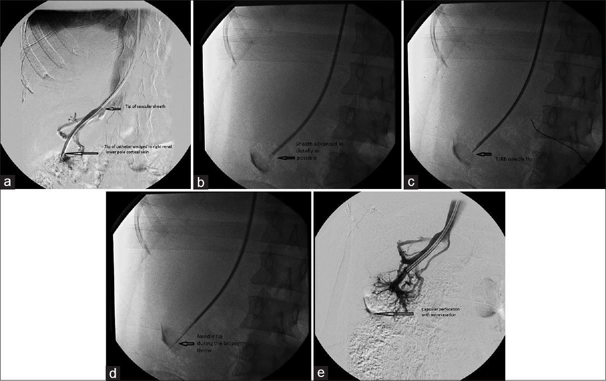
- Sequential images in a patient undergoing transjugular renal biopsy. Right renal venogram (a) with tip of vascular sheath in the proximal right renal vein through which catheter is wedged distally into the right lower pole renal cortical vein. Following the venogram, the sheath is advanced as distally as possible into the cortical vein to allow introduction of transjugular renal biopsy needle (b). Renal access and biopsy set needle was advanced through the sheath and placed distally into peripheral cortical vein (c) and the biopsy was performed (d) postbiopsy check run (e) shows capsular extravasation
All patients returned to the ward for routine 24-h bed rest and hemodynamic observation, which took place every 15 min for the first 2 h, then half-hourly for 4 h, and then hourly. Follow-up was done with USG scan 3 h after the procedure and on day 3 [Figure 2].

- Follow-up ultrasonogram on the 3rd day postbiopsy shows no perinephric collection, renal arteriovenous fistula, or pseudoaneurysm. Clinically, patient had no hematuria
Tissue adequacy
The number of needle passes and cores obtained per procedure were ascertained. The tissue cores were considered adequate when a sufficient number of intact glomeruli were present to make a pathological diagnosis.
Results
There were 8 males and 1 female, with a mean age of 41.5 ± 15.4 years. The medical reasons for renal biopsy and the clinical parameters for nine patients are summarized in Table 1. These included patients with recent deterioration in renal function, patients with multiple comorbidities investigated for chronic renal failure, and new presentations of nephrotic syndrome or significant proteinuria. All patients were referred by nephrologists who requested renal biopsy specifically via the transjugular route, due to relative contraindications to percutaneous biopsy. Six of the nine patients (66.67%) had a platelet count of <50,000/mcL, 2 (33.3%) had an elevated INR of more than 1.4, and 1 had both.

The right IJV access was achieved in all the nine patients, and biopsy specimens were obtained from the right kidney in all cases. A total of 3.2 ± 0.4 cores was obtained, with median (range) number of glomeruli being 10 (7–11) at light microscopy. Adequate renal tissue was obtained in all the patients as confirmed by the pathologists. The mean duration of the procedure was 50 min (range, 30–120 min) including the time for transferring the patient to and from the couch in the interventional radiology room. The amount of contrast material used to verify location before biopsy was 11.8 ± 2.3 ml.
Table 2 summarizes the results of TJRB in our series.

Complications
Although capsular perforation was a consequence in 5 out of 9 patients, none of them had major complication requiring management (either endovascular treatment or blood transfusion).
Discussion
Percutaneous renal biopsy is a commonly performed safe procedure, with an excellent yield, ranging from 95.5% to 98.8% in the published literature,[101112] and it is the routine method of acquiring renal tissue in patients. The main risk is bleeding due to the high vascularity of the kidneys. Since the diseased kidneys are often small with cortical thinning, the tamponade effect may be minimal. In addition, early clinical detection of retroperitoneal hemorrhage is difficult. In patients in whom a percutaneous biopsy is contraindicated, when the pathological diagnosis alters clinical management, TJRB provides an alternative approach. Though the procedure is utilized commonly in the Western world with published data showing its safety and efficacy using both the aspiration[7101314] and the core biopsy[1516171819] techniques, it has still not gained popularity in the Indian subcontinent despite its potential to open possibilities for the management of a significant subgroup of patients who are considered high risk for both percutaneous and open kidney biopsy. Considering the cost of the procedure (approximately INR 40,000 at our center including the cost of hardware which is approximately INR 35,000), it has the potential for widespread application.
Patients were referred to us for TJRB with varying degrees of renal impairment, proteinuria, and/or hematuria as illustrated in Table 1. Many of them had significant comorbidities, necessitating a biopsy to decide on the optimal clinical management. All patients had bleeding diathesis (thrombocytopenia and/or coagulopathy) which contraindicated percutaneous biopsy. Other recognized indications for a TJRB include a single functioning kidney, horseshoe kidney, end-stage renal disease or small kidneys, and patients' inability to lie in the prone position such as those with a particularly high BMI, uncooperative patients, patients in Intensive Care Unit with respiratory assistance, and patients with voluminous ascites.[20] In patients with acute renal failure requiring hemodialysis, TJRB can be usefully combined with central venous dialysis catheter placement.[20] Concomitant TJRB can be performed in conjunction with transjugular liver biopsy in patients undergoing assessment for potential liver transplantation, to differentiate between hepatorenal syndrome and other renal lesions that may progress.[212223]
A lower pole renal vein is preferred for biopsy due to optimal angle for cannulation. Performing the biopsy peripherally also reduces the chances of damaging a large vessel. The low venous pressure and direction of venous flow also reduce the incidence of severe hemorrhage, unless inadvertent arterial puncture occurs. TJRB may be performed using an aspiration needle or core biopsy system. A diagnostic yield of 73–95% and major complication rate of 1–18%[791013] have been reported using the aspiration needle, compared with the yields of 89–96.5% and major complications of 2.7–27% with the core biopsy needle.[15161718] Diagnosis is dependent on adequate cortical sampling, providing a sufficient number of glomeruli. The median number of glomeruli per patient of ten for light microscopy in our series is comparable to the 9.8–10.8 in the aspiration series[7101314] and 9–9.8 reported in the core biopsy series.[1618] The samples were adequate for pathological diagnosis in all patients.
Like most interventional procedures, the number of biopsy attempts is largely dependent on the favorability of the anatomy and operator experience. At our institution, the procedure is performed by an interventional radiologist. The average number of needle passes in our series was 5.3, which is comparable to those of 4–5.5 in other studies.[1617] The mean number of core specimens of 3 ± 1 is also comparable to those in the literature.[1618] In the published series using the core biopsy system,[15161718] there was no mention of a limit to the number of needle passes. In two published series using the aspiration technique, the number of passes was limited to a maximum of 3 and 8, respectively, depending on the patients' risk factors.[710] However, increasing the number of biopsies does not necessarily increase the tissue yield, as cortical tissue (in entirety or mixed with medulla or fat) is more likely to be obtained following the first and second needle passes. The yield deteriorates significantly with subsequent needle passes.[24] This suggests that the first needle pass is the most important and that subsequent biopsies may yield fragmented or crushed specimens, making accurate diagnosis more difficult. Therefore, in patients with bleeding diathesis, perhaps, it is reasonable to restrict the number of passes to three attempts. Moreover, it has been shown that six or more needle passes have a higher incidence of capsular perforation compared to five or fewer passes.[19]
The association between capsular perforation and TJRB is well recognized and this has not been considered a complication per se.[1011] Of the nine TJRB procedures in our series, five (55.56%) were associated with capsular perforation. None had any clinical sequelae or required blood transfusion. Capsular or subcapsular perforation should theoretically indicate that the biopsy included cortex and was likely to allow a diagnosis to be made. Elective coil embolization of the biopsy track was performed in five of the early cases of isolated capsular perforation by See et al.[24] However, isolated capsular/subcapsular extravasation is mainly subclinical, and elective embolization of the biopsy track is probably not indicated in the majority of cases, unless the patient is hemodynamically unstable. There is no published evidence on the benefit of elective embolization of the biopsy track following capsular perforation.
Collecting system puncture at the time of TJRB was not noted in any patient in our series but occurred in five cases, with or without concurrent pericapsular extravasation in the study by See et al. where in a further four cases, subsequent macrohematuria implied that the collecting system was breached.[24] The reported rates of major complications (which included the need for postprocedure blood transfusion) using the aspiration biopsy techniques range from 1% to 18%,[7101314] compared with 2.7–27% for the core biopsy system.[15161718] In our series, there were no major complications. Contrast-induced renal failure is a concern, given that many patients already have some degree of renal impairment. However, only a small volume of contrast medium (15–30 ml) is usually required, and this is unlikely to be of significance. Routine postprocedure ultrasonography has not been recommended,[19] but in our series all patients underwent ultrasonography 3 h after the procedure and on day 3. Persistent severe loin pain, frank hematuria, and clot retention will necessitate sonography. Other potential but rare complications of the transjugular route include arrhythmias, pneumothorax, and hemothorax.
Our initial experience with TJRB is similar or better than in prior reports with regard to both diagnostic yield and complication rates.[10] This difference may be related to the small sample size. The best tissue yields are obtained with the first two needle passes. In patients with a bleeding diathesis, it seems prudent to limit the number of needle passes to no more than three.
Table 3 provides a comparison of the technical adequacy and complications encountered in our series against case series by Abbott et al. and See et al., as well as studies by Cluzel et al. and Sofocleous et al.

In summary, TJRB is recognized as an alternative, safe, and effective technique in patients with renal parenchymal disease when contraindications to the percutaneous approach exist.
Conclusion
TJRB is a safe procedure in patients with contraindications to percutaneous biopsy and is associated with only a low risk of major complications.
Financial support and sponsorship
Nil.
Conflicts of interest
There are no conflicts of interest.
References
- Percutaneous native renal biopsy: Comparison of a 1.2-mm spring-driven system with a traditional 2-mm hand-driven system. Am J Kidney Dis. 1994;23:498-503.
- [Google Scholar]
- Clinical utility of kidney biopsies in the diagnosis and management of renal disease. Am J Nephrol. 1989;9:309-15.
- [Google Scholar]
- Controversy: The role of renal biopsy in modern medicine. Am J Kidney Dis. 1982;1:241-3.
- [Google Scholar]
- Transvenous renal biopsy: A preliminary report on 36 patients (abstr) Kidney Int. 1990;37:279.
- [Google Scholar]
- The diagnostic yield of transjugular renal biopsy. Experience in 200 cases. Kidney Int. 1992;41:445-9.
- [Google Scholar]
- Initial experience with the transjugular renal biopsy. Cas Lek Cesk. 1993;132:219-20.
- [Google Scholar]
- Transjugular versus percutaneous renal biopsy for the diagnosis of parenchymal disease: Comparison of sampling effectiveness and complications. Radiology. 2000;215:689-93.
- [Google Scholar]
- Safety of ultrasound-guided percutaneous renal biopsy-retrospective analysis of 1090 consecutive cases. Nephrol Dial Transplant. 1998;13:975-7.
- [Google Scholar]
- Percutaneous biopsy in diffuse renal disease: Comparison of 18- and 14-gauge automated biopsy devices. J Vasc Interv Radiol. 1998;9:651-5.
- [Google Scholar]
- Transjugular renal biopsy in the treatment of patients with cirrhosis and renal abnormalities. Hepatology. 1996;24:1143-7.
- [Google Scholar]
- Transjugular renal biopsy. Our experience with 67 cases. Kidney Blood Press Res. 2001;24:207-12.
- [Google Scholar]
- Transjugular renal biopsy in high-risk patients: An American case series. BMC Nephrol. 2002;3:5.
- [Google Scholar]
- Diagnostic utility and safety of transjugular kidney biopsy in the obese patient. Nephrol Dial Transplant. 2004;19:1798-802.
- [Google Scholar]
- Transjugular renal biopsy in patients with liver disease. Am J Kidney Dis. 2001;37:1144-51.
- [Google Scholar]
- Transvenous transjugular renal core biopsy with a redesigned biopsy set including a blunt-tipped needle. Cardiovasc Intervent Radiol. 2002;25:155-7.
- [Google Scholar]
- Renal biopsy in high-risk patients with medical diseases of the kidney. Am J Kidney Dis. 2000;36:419-33.
- [Google Scholar]
- The current spectrum of infectious glomerulonephritis. Experience with 76 patients and review of the literature. Medicine (Baltimore). 1995;74:63-73.
- [Google Scholar]
- Contribution of transjugular liver biopsy in patients with the clinical presentation of acute liver failure. Cardiovasc Intervent Radiol. 2006;29:1008-10.
- [Google Scholar]
- Transjugular renal biopsy: Our experience and technical considerations. Cardiovasc Intervent Radiol. 2008;31:906-18.
- [Google Scholar]


