

Translate this page into:
Brachial plexus compression due to subclavian artery pseudoaneurysm from internal jugular vein catheterization
This is an open access article distributed under the terms of the Creative Commons Attribution-NonCommercial-ShareAlike 3.0 License, which allows others to remix, tweak, and build upon the work non-commercially, as long as the author is credited and the new creations are licensed under the identical terms.
This article was originally published by Medknow Publications & Media Pvt Ltd and was migrated to Scientific Scholar after the change of Publisher.
Abstract
Internal jugular vein (IJV) catheterization has become the preferred approach for temporary vascular access for hemodialysis. However, complications such as internal carotid artery puncture, vessel erosion, thrombosis, and infection may occur. We report a case of brachial plexus palsy due to compression by right subclavian artery pseudoaneurysm as a result of IJV catheterization in a patient who was under maintenance hemodialysis.
Keywords
Brachial plexus palsy
internal jugular vein catheterization
pseudoaneurysm
subclavian artery

Introduction
Internal jugular vein (IJV) catheterization is commonly used to obtain temporary access to circulation enabling hemodialysis. However, significant complications such as internal carotid artery (ICA) puncture, pneumothorax, vessel erosion, thrombosis, airway obstruction, and infection can occur. The most common complication is ICA puncture with incidence of 9.3%.[1] Hemodialysis patients may have to undergo multiple catheter placements and vascular access interventions. This, along with their comorbid conditions, increases the risk of such complications.[123] Here, we report a patient on hemodialysis who developed right subclavian artery pseudoaneurysm following the right IJV catheterization. Jugular venous catheterization is generally safer than subclavian venous catheterization. Jugular vein, therefore, has become the preferred site for hemodialysis catheter insertion. We are describing a case of brachial plexus compression attributable to delayed recognition of a right subclavian artery pseudoaneurysm as a complication of jugular venous catheterization of hemodialysis catheter. Any neck swelling, new bruit, and the symptoms of brachial plexopathy after jugular venous catheterization warrant an intensive investigation to exclude arterial injury.
Case Report
A 20-year-old boy with end-stage renal disease was admitted to a hospital with metabolic acidosis and uremia. An 18 gauge needle was inserted percutaneously by landmark method via central approach which resulted in arterial puncture; needle was removed, and firm pressure was applied for 5 min. The second attempt via low central approach was done so as to avoid carotid artery puncture which was successful and a double lumen hemodialysis catheter was inserted with Seldinger technique via right IJV, and the patient underwent heparin free hemodialysis. The patient complained of pain and swelling in the right supraclavicular region after 4 days of insertion of the catheter and then the swelling increased in size along with weakness in his right upper limb. Brachial compression due to hematoma was suspected. Color Doppler study of neck vessels showed a partially thrombosed pseudoaneurysm formation of size 5.6 cm × 4.2 cm seen in the right supraclavicular region, showing underlying aneurysm measuring 1.9 cm × 1.3 cm within the lesion which showed turbulent flow with whirling in the pseudoaneurysm and appeared to be communicating with right subclavian artery with normal color flow in carotid artery and IJV. The patient was subjected to computed tomography angiography to determine origin of pseudoaneurysm which revealed 5.5 cm × 7.5 cm × 8.5 cm hematoma in right subclavian fossa with blood fluid levels [Figures 1 and 2]. There was extravasation of contrast into hematoma likely from thyrocervical trunk approximately 7.5 mm away from its origin from subclavian artery. Over next 24 h, the patient was unable to move his right upper limb with increasing pain, so he was subjected to open surgical pseudoaneurysmectomy.
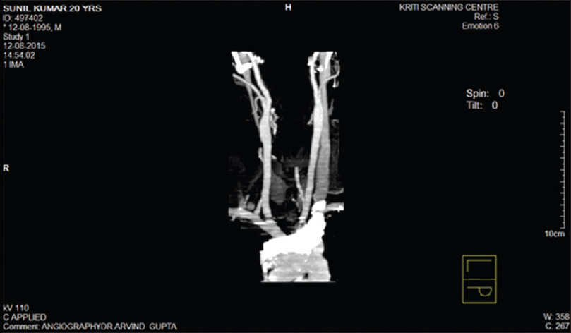
- Computed tomography angiography of neck vessels

- Computed tomography angiography of neck vessels showing pseudoaneurysm on the right side
Discussion
Right IJV catheterization is commonly used perioperatively for invasive monitoring as well as administration of fluid and vasoactive drugs and emergent hemodialysis. The advantages are straight course into superior vena cava, superficial location, and definite landmarks for placement. Arterial injury leading to hematoma formation, arterial dissection, arteriovenous fistula, or pseudoaneurysm is known as catheter-related cervicothoracic arterial injuries. Mallory et al. published a prospective, randomized study indicating a higher rate of success and a lower number of attempts and immediate complications for IJV catheterization with bi-dimensional ultrasound versus the anatomical landmarks technique[2] Dolu et al. also found that IJV catheterization guided by real-time ultrasonography (USG) resulted in a lower access time and lower rate of attempts.[3]
Pseudoaneurysm results from a variety of causes such as infection, trauma, and surgical procedures. The most common mechanism is disruption of arterial continuity with extravasation of blood into surrounding tissue. This results in the formation of fibrous tissue capsule which progressively enlarges because of underlying arterial pressure.[4] In the present case, the accidental penetration injury of the right subclavian artery associated with low puncture might have led to pseudoaneurysm formation which progressively expanded due to arterial pressure.[5]
Differentiation between simple hematoma and pseudoaneurysm may be difficult by clinical examination alone. Hematoma usually appears shortly after the procedure and tend to resolve in time depending on size, location, and extent of injury whereas pseudoaneurysm may appear later with pulsatile and expanding mass. Duplex USG will help to differentiate between two, and a selective angiogram is necessary to determine precise origin and extent of injury.[6] In our case, initially, we attributed brachial plexus compression due to hematoma in neck.
Because of close anatomic relationship between brachial plexus and subclavian artery in the thoracic inlet, even a small false aneurysm can result in compression injury to the neuroplexus.[78] Because brachial palsy has a poor prognosis when recognition is delayed, an aggressive approach is advocated. In our review, early surgical intervention of compressive hematoma within 48 h resulted in improvement in all patients while late intervention after 48 h resulted in improvement in about half of patients.[78910] Our patient had progressive signs and symptoms of brachial plexopathy.
Treatment options for pseudoaneurysm are USG-guided compression, percutaneous thrombin injection, coil embolization, endovascular stents, and open surgical repair.[111213] USG-guided compression which is frequently used for ablation of femoral pseudoaneurysm was not possible in our case because of depth of the artery and overlying bone. Cardiothoracic and vascular surgeon was consulted for endovascular stenting but was not feasible due to risk for cerebral embolization and stroke. Thus, open surgical repair was done. Few studies showed that early intervention within 48 h resulted in improvement in all patients while late intervention resulted in improvement in about half of patients.[78910] Our patient had only partial improvement.
Conclusion
For all traumatic injuries of attempted jugular venous catheterization, particularly arterial puncture, an aggressive investigational approach is recommended. Any neck swelling or symptoms of brachial plexopathy should arise the suspicion of pseudoaneurysm and confirm by color Doppler study. A symptomatic pseudoaneurysm should be treated without delay to prevent permanent neurological damage.
Financial support and sponsorship
Nil.
Conflicts of interest
There are no conflicts of interest.
References
- Brachial plexus palsy due to subclavian artery pseudo aneurysm from internal jugular catheterization. Indian J Crit Care Med. 2007;11:93-5.
- [Google Scholar]
- Ultrasound guidance improves the success rate of internal jugular vein cannulation. A prospective, randomized trial. Chest. 1990;98:157-60.
- [Google Scholar]
- Comparison of an ultrasound-guided technique versus a landmark-guided technique for internal jugular vein cannulation. J Clin Monit Comput. 2015;29:177-82.
- [Google Scholar]
- Pseudoaneurysms. In: Rutherford RB, ed. Vascular Surgery (4th ed). Philadelphia: W B Saunders; 1995. p. :1153-61.
- [Google Scholar]
- Arterial injuries following diagnostic, therapeutic, and accidental arterial cannulation in haemodialysis patients. Nephrol Dial Transplant. 1997;12:1448-52.
- [Google Scholar]
- Surgery: Scientific Principles and Practice. Philadelphia: Lippincott; 1993. p. :1683.
- Brachial plexus compression: Complication of delayed recognition of arterial injuries of the shoulder girdle. Arch Surg. 1981;116:175-8.
- [Google Scholar]
- Subclavian artery false aneurysm associated with brachial plexus palsy: A complication of parenteral drug addiction. Am J Emerg Med. 1990;8:129-33.
- [Google Scholar]
- Delayed brachial plexus paralysis due to subclavian pseudoaneurysm after clavicular fracture. Eur J Cardiothorac Surg. 1993;7:497-8.
- [Google Scholar]
- Thrombin injection for treating a subclavian artery pseudoaneurysm. Surgery. 2000;127:716-8.
- [Google Scholar]
- Endovascular stented graft repair of a pseudoaneurysm of the subclavian artery caused by percutaneous internal jugular vein cannulation: Case report. Am J Crit Care. 1995;4:472-5.
- [Google Scholar]
- Transluminally placed endovascular stented graft repair for arterial trauma. J Vasc Surg. 1994;20:466-72.
- [Google Scholar]


