

Translate this page into:
Right Renal Biopsy Through Petit's triangle
-
Received: ,
Accepted: ,
This is an open access journal, and articles are distributed under the terms of the Creative Commons Attribution-NonCommercial-ShareAlike 4.0 License, which allows others to remix, tweak, and build upon the work non-commercially, as long as appropriate credit is given and the new creations are licensed under the identical terms.
This article was originally published by Wolters Kluwer - Medknow and was migrated to Scientific Scholar after the change of Publisher.
Sir,
Per-cutaneous renal biopsy is usually performed in the prone position, and the preferred site of biopsy is the lower pole of the left kidney.[1] Prone position may not be feasible for patients who have abdominal distension due to obesity, ascites or pregnancy, and in whom such posture might provoke or aggravate respiratory distress. A new patient positioning method namely the supine antero lateral position (SALP) was described in 2008 by Gesualdo et al.,[2] that facilitated per-cutaneous renal biopsy in obese patients and patients with respiratory difficulty. We present the methodology for performing per-cutaneous biopsy from right kidney with patient lying in left lateral position.
A 70-year-old gentleman presented with nephrotic syndrome with massive proteinuria (12 gm/24 hours), normal renal function, and anasarca. His BP was 160/100 and body mass index (BMI) was 29. He had bilateral pleural effusion and tense ascites. He was treated with diuretics for a week, nevertheless ascites persisted. Renal biopsy was indicated and routine evaluation for per-cutaneous renal biopsy was done. Patient was unable to assume prone position and hence renal biopsy had to be performed with patient lying in a different position. [Figure 1a] reveals the patient positioning for obtaining per-cutaneous right renal biopsy in this patient. The patient lied in the left lateral position with the torso lying perpendicular to the horizontal plane and the right upper limb placed way from right flank. The left lateral position was chosen since the patient felt comfortable with this position and it provided a clear window to visualize the right kidney between the right lower costal border and the iliac crest with ultrasound scan. The right kidney was visualized with ultrasound and a “safe path” for passing the needle was identified [Figure 1b]. The safe path was the zone in which no part of other organ like liver or colon was intervening between the skin and the kidney. Under local anesthesia ultrasound guided per-cutaneous biopsy was taken from right kidney by passing an 18-gauge automatic spring-loaded biopsy gun through the safe path, which was identified between the lower costal border and iliac crest. Two passes were made to obtain two cores of renal tissue. The total procedural duration including scanning, draping, and biopsy was 15 min. Patient was comfortable throughout the procedure. There was no post biopsy complication like hematuria, perinephric hematoma, or inadvertent injury to liver or other intra-abdominal organs. There was no ascitic leak. The samples were adequate in size for interpreting the histology and making a diagnosis.
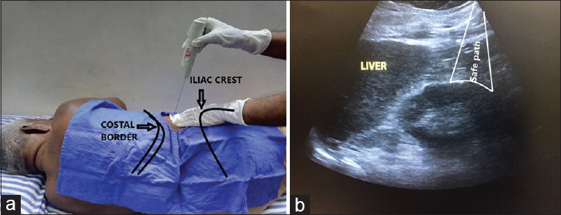
- (a) Patient positioning and landmarks for obtaining right renal biopsy in left lateral position. (b) "Safe path" identified by ultrasound for passing needle
In the position described by Gesualdo et al., the SALP was obtained by placing towels underneath the ipsilateral shoulder and gluteus, which elevated the flank by 30°.[2] The SALP exposes the Petit's triangle, which is bounded anteriorly by external oblique, posteriorly by latismus dorsi, and inferiorly by iliac crest.[3] The Petit's triangle provides an easy access to the lower pole of the kidney while performing per-cutaneous renal biopsy. The SALP may be regarded as a manoeuvre to expose the Petit's triangle in obese individuals.[2] In this case the patient was placed in the left lateral position with torso lying perpendicular to the horizontal plane and the Petit's triangle was apparent, which enabled biopsy in the left lateral position.
Conclusion
The lateral decubitus position is a simple and effective method for obtaining per-cutaneous renal biopsy in patients who cannot lie prone because of abdominal distention.
Declaration of patient consent
The author certifies that he has obtained all appropriate patient consent forms. In the form the patient has given his consent for his images and other clinical information to be reported in the journal. The patient understands that his name and initials will not be published and due efforts will be made to conceal his identity, but anonymity cannot be guaranteed.
Financial support and sponsorship
Nil.
Conflicts of interest
There are no conflicts of interest.
References
- Basics of kidney biopsy: A nephrologist's perspective. Indian J Nephrol. 2013;23:243-52.
- [Google Scholar]
- Percutaneous ultrasound-guided renal biopsy in supine antero-lateral position: A new approach for obese and non-obese patients. Nephrol Dial Transplant. 2008;23:971-6.
- [Google Scholar]
- Clinical relevance of Petit'striangle: A forgotten landmark. Rev Urol. 2018;20:112-4.
- [Google Scholar]


